Inhibition
Electrical stimulation of the caudate nucleus can cause a variety of inhibitory effects, including inhibition of aggressiveness, motor arrest, sleep, adynamia (*), food refusal, and amnesia.
An inactive state accompanied by loss of muscle tone.
It has also been shown that electrical stimulation of the septal area can inhibit aggression, and that of the claustrum can produce disruption of consciousness.
If abused, this would make it possible to inhibit people who are angry or dissatisfied with injustice and forestall rallies, demonstrations, and protests.
At least in fact, an experiment was already conducted more than 50 years ago in which the brain of a chimpanzee was wirelessly connected to a computer, and when excited states were detected in the brainwaves, the brain was remotely stimulated to make it docile.
Table of ContentsAll_Pages
Circuitry
Caudate Nucleus
The basal ganglia is a neuronal circuit necessary for voluntary movement control, and the striatum is the input module of it. (Báez-Mendoza and Schultz 2013)
The striatum is composed of three nuclei: the caudate nucleus, the putamen, and the ventral striatum/nucleus accumbens.
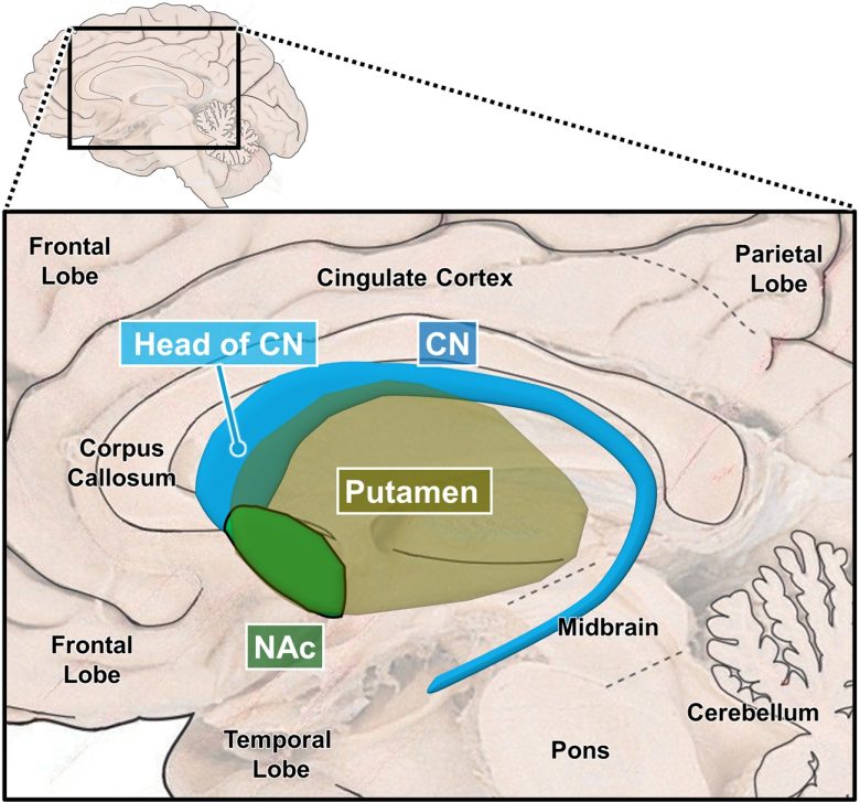
Structure of the Striatum
(The brain image from Vanderah 2018)
CN = caudate nucleus; NAc = nucleus accumbens.
Although the functional role of the caudate nucleus is in general explained as a component of the neural circuitry of the basal ganglia, some researchers explain it in terms of an independent nucleus with its own function. (Villablanca 2011)
Damage to the caudate nucleus is known to cause humans and animals to become impulsive, exhibit compulsive behavior, and lose inhibition.
Cats with both of the caudate nuclei removed persistently approached and then followed a person, another cat, or any object, and attempted to contact them. In addition, the cat became more awake and showed hyposomnia.
A monkey with both of the caudate nuclei removed showed hyperactivity, pacing back and forth in the cage compulsively. Weeks later the pacing changed into circling the cage.
A woman who had both of the heads of the caudate nuclei damaged displayed abnormal behavior inappropriate for her previous condition. (Richfield et al. 1987)
These were mostly symptoms of disinhibition, including vulgarity, impulsiveness, easy frustration, violent outbursts, polydipsia and enuresis, wandering, increased appetite, hypersexuality, and minor criminal behavior including shoplifting and exposing herself, and poor hygiene.
When comparing the head of the caudate nucleus in children with attention deficit hyperactivity disorder (ADHD) with that in normal children, the left side was found to be significantly smaller. (Hynd et al. 1993)
On the contrary, electrical stimulation of the caudate nucleus produces the effects exactly opposite to the disinhibition caused by lesions: inhibition.
Electrical stimulation of the caudate nucleus, particularly the head of the caudate nucleus, has been shown to inhibit aggression and appetite, to arrest ongoing movements, and to induce sleep and adynamia.
Septal Area, Claustrum
Patients with tumors in the septal area exhibited aggression outbursts and increased irritability, known as "septal rage," and rodent studies have supported this observation. (Menon et al. 2022)
Conversely, electrical stimulation of the septal area has been shown to inhibit aggression.
The claustrum synchronizes inhibitory neurons in the widespread neocortical areas, and is involved in generating slow waves, the brainwaves seen during sleep and rest. (Narikiyo et al. 2020)
Electrical stimulation of the claustrum has been shown to cause disruption of consciousness in humans and to produce a similar state in animals.
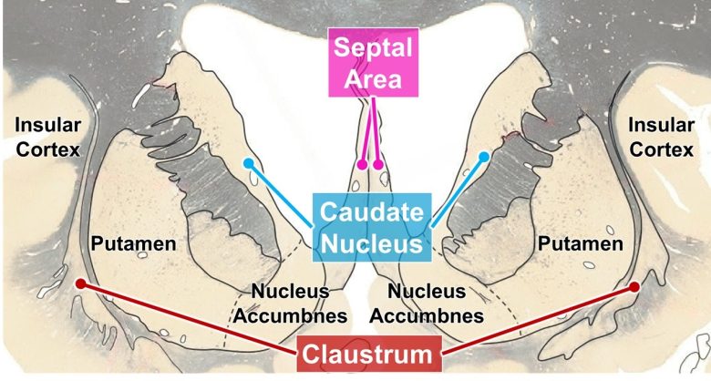
Location of the caudate nucleus, septal area, and claustrum
(The brain image from Vanderah 2018)
Inhibition of Aggressiveness
Electrical stimulation of the caudate nucleus and septal area has been shown to inhibit aggression in animals, such as monkeys.
Delgado's Animal Experiments
Jose Delgado, a Spanish neurophysiologist at Yale University, invented a wireless brain stimulation device, which he called the stimoceiver.
He attached the device to rhesus monkeys, gibbons, chimpanzees, and bulls, conducting numerous experiments aimed at inhibition of aggressiveness.
Bulls
The most famous one is an experiment in which Delgado himself confronted a bull and stopped its violent charge by remote control. (Marzullo 2017)
The experiment was done on a ranch in Córdoba, Spain, in 1964.
The bull "Lucero" had a stimoceiver attached, allowing for remote stimulation of the caudate nucleus, thalamus, and primary motor cortex via radio waves.
Lucero continued demonstrating robust charging behavior after the surgery.
When the bull was charging at a bullfighter, electrical stimulation of the caudate nucleus or thalamus was delivered at 1 mA, and then the bull stopped immediately.
Lucero was still during the stimulation, although blinking its eyes and breathing regularly.
The bull returned furiously to a full charge when the electrical stimulation ceased.

Next, Delgado, armed with a radio transmitter, confronted the bull himself.
When Lucero was charging Delgado and had reached within 2 or 3 meters of him, Delgado turned on the stimulation.
And the bull stopped violently, causing a cloud of dust with his legs halting suddenly, his tail lowering, and his head raising, and maintaining a state of disappeared aggressiveness.
Delgado then backed away, sought shelter behind a wooden barrier in the ring, and turned off the stimulation, and the bull immediately charged again and rammed the barrier.

In another bull named “Cayetano,” electrical stimulation of the left caudate nucleus induced rotational movements to the left.
With the stimulation at 0.5 mA, the bull moved its head to the left, and at 0.7 mA, the bull moved its head to the left and spun in a complete circle.
When the stimulation was applied to the right caudate nucleus, similar behavior was observed, but the bull turned in the opposite direction, i.e., to the right.
Stimulation of the caudate nucleus is known to produce rotational movements as well as general inhibition.
Chimpanzees
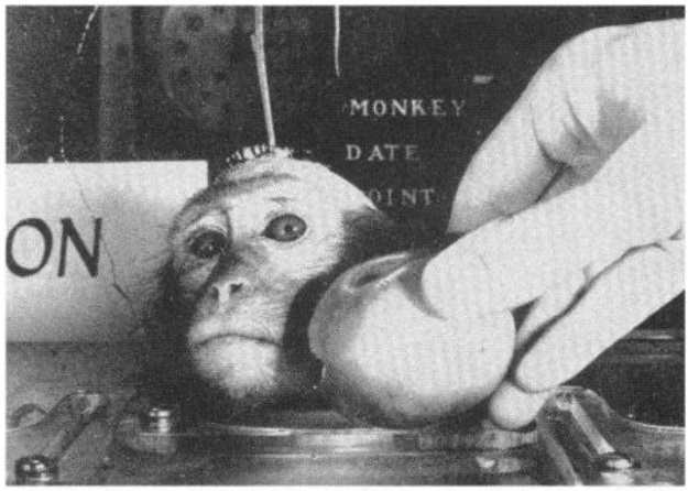
Another famous one is an experiment in which he wirelessly connected the brain of a chimpanzee to a computer, which automatically detected a state of excitement and sent pain signals, ultimately rendering him lethargic. (Delgado et al. 1970)
The purpose of this experiment says "to demonstrate the principle that telemetered brainwaves can be pattern recognized by a computer in order to trigger contingent radio-stimulation of a determined area of the brain."
It is obvious that this was an experiment for military purposes, with applications such as crowd control in mind, and indeed, this experiment was conducted within a U.S. Air Force military facility.
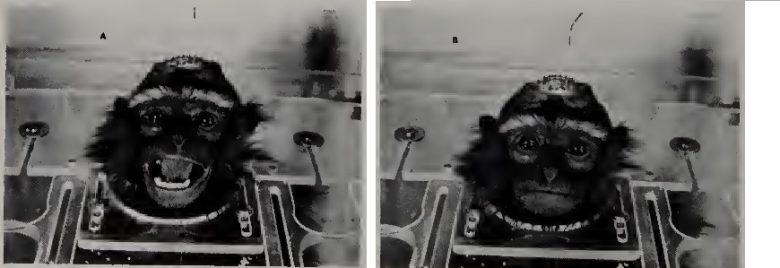
The subject was a six-year-old juvenile male chimpanzee named "Paddy."
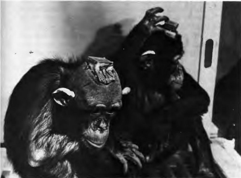
Two chimpanzees with the stimoceiver attached. Paddy on the left and Carlos on the right. (Delgado 1970)
Paddy had a stimoceiver attached to his brain and electrodes implanted in the amygdala and the reticular formation.
Brainwaves of the amygdala were wirelessly transmitted and analyzed by a computer in the laboratory. When spindles were detected, a stimulation signal to the reticular formation was automatically delivered.
Amygdaloid spindles were associated with excitement in the chimpanzee, and electrical stimulation of the reticular formation caused pain in Paddy.
That is, a system was established where the computer automatically detects when Paddy becomes excited and sends pain signals to his brain.

Paddy had been kept free-roaming with other chimpanzees on an artificial island built on a U.S. Air Force base.
Upon initiating the system operation, Paddy's amygdala spindles were reduced by half within two hours and almost disappeared within six days.
Ten days after the operation, a visual and olfactory discrimination task was administered, which was carried out without error.
There was, however, an important behavioral change.
Paddy sat very quietly, performing the tasks without observing the food pellets being delivered and often without eating them.
The lack of attention and motivation was evident, and the attitude of Paddy was very different from his usual behavior before the experiment.
He was more placid than usual, without being excited by the presence of visitors or by the offering of appetizing food, ending up in a state of lethargy.
Delgado also showed more directly that electrical stimulation of the caudate nucleus can inhibit aggression in chimpanzees. (Delgado 1970)
The subject "Carlos" was, like most chimpanzees, rather temperamental and was easily provoked into a tantrum by being punished, frustrated, or merely teased.
He liked to be touched by people he knew but not by strangers, and showed a defensive, anxious reaction when approached by an unfamiliar investigator.
Electrical stimulation of the head of the caudate nucleus inhibited these defensive attitudes.
He displayed no emotion, appeared peaceful, and could be teased without any resulting disturbance.

Rhesus Monkeys
Delgado's experiments on inhibition of aggressiveness through electrical brain stimulation primarily used rhesus monkeys. (Delgado 1964)
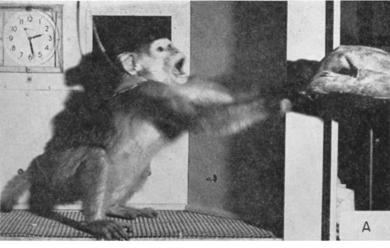
Rhesus monkeys are ferocious animals.
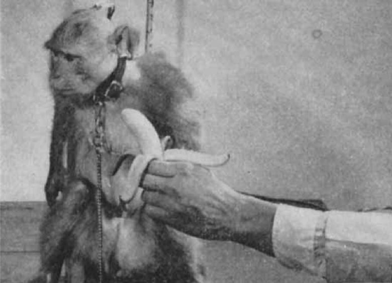
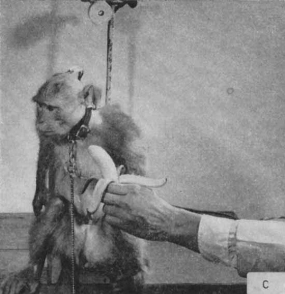
If a glove, a stick, or the experimenter’s hand suddenly approached a monkey, it usually opened its mouth, showing teeth, moved its head forward, vocalized with a characteristic low-toned sound, and tried to grab, to scratch, and to bite.
Stimulation of some points of the caudate nucleus made the monkey close its mouth and lose its aggressive attitude.
It was then safe to touch the monkey, and even to put a finger in its mouth. When a glove or stick came near its face, the monkey used its hand to push it away without attacking.
As soon as the caudate stimulation stopped, the monkeys reverted to their usual ferocious behavior.

Delgado conducted an experiment to see how electrical brain stimulation affected the behavior of monkeys in colonies. (Delgado 1963)
In the colony of four rhesus monkeys, electrodes were placed in various sites of the brains of the boss male monkey, "Ali," and the second-ranking female monkey, "Sarah."
In both, electrical stimulation of the periaqueductal gray (PAG) induced antisocial behavior with increased chasing, jumping, biting, and fighting.
On the contrary, electrical stimulation of the head of the caudate nucleus reduced spontaneous aggressiveness. This effect was more impressive in Ali because of his greater size and ferocity.
It also produced loss of interest in food, and inhibition of various activities, such as drinking, taking of pellets, walking, picking, and self-grooming.
During caudate stimulation it was possible to touch the monkey with one's bare hands, and in this manner the monkeys were caught repeatedly.
Next, Delgado placed a lever in the colony that delivers electrical stimulation of the head of the caudate nucleus of the boss monkey Ali, and observed how the subordinate monkeys behaved.
The third-ranking female monkey, "Elsa," who had a hostile relationship with Ali, was observed several times to press the lever and reduce Ali's aggressiveness when she was threatened.
Her attention was directed not to the lever but to Ali while pressing it. It was unusual behavior because if lower-ranking monkeys look straight at the boss of the colony, they will be retaliated against.

Delgado also showed that electrical stimulation of the septal area can inhibit aggression. (Rubinstein and Delgado 1963)
Threatening a restrained monkey with a leather glove elicited an aggressive display against the experimenter.
The monkey showed its teeth, tried to grab and pull the glove, vocalized loudly, and bit the glove ferociously when possible.
Electrical stimulation of the septal area inhibited these aggressive responses.
The monkey merely looked at the approaching glove without signs of excitement such as vocalization or biting. However, it turned its head to one side and often tried to push the glove slowly away.
During stimulation it was safe to touch the monkey’s face or mouth with bare hands.
Gibbons
Delgado also conducted experiments using gibbons on inhibition of aggressiveness. (Delgado 1975)
With a gibbon placed in a restraint chair, stroking its face lightly with a leather glove usually induced an aggressive reaction consisting of showing the teeth, attempts to grab and pull the glove, vocalization, and biting.
During electrical stimulation of the caudate nucleus, however, the gibbon looked at the approaching glove without signs of excitement or hostility, and it was then safe to touch its mouth.
Electrical stimulation of the head of the caudate nucleus also inhibited aggression induced by electrical stimulation of other sites of the brain.
In a gibbon restrained in a chair, stimulation of the PAG produced aggressive behavior.
This was manifested in restlessness, showing of the teeth, attempts to grab and bite, and vocalizations.
When electrical stimulation of the caudate nucleus preceded the PAG stimulation by 2 seconds, the stimulation-induced aggressive behavior disappeared.
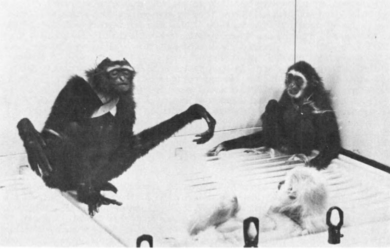
Similar results were obtained with gibbons free in a colony cage.
Following electrical stimulation of the PAG, black gibbon "Gaily" launched a well-directed attack against blonde gibbon "Coti."
Coti defended itself and retaliated, in spite of its inferior size and strength. Usually a brief 2-second stimulation induced threats and fights which continued for 10-60 seconds.
But by electrical stimulation of the caudate nucleus, Gaily was immediately inhibited, stopped the fighting, and assumed an innocent and peaceful posture.

Other Animal Experiments
The inhibition of aggressiveness by electrical stimulation of the caudate nucleus and septal area has been confirmed by several researchers.
Cats
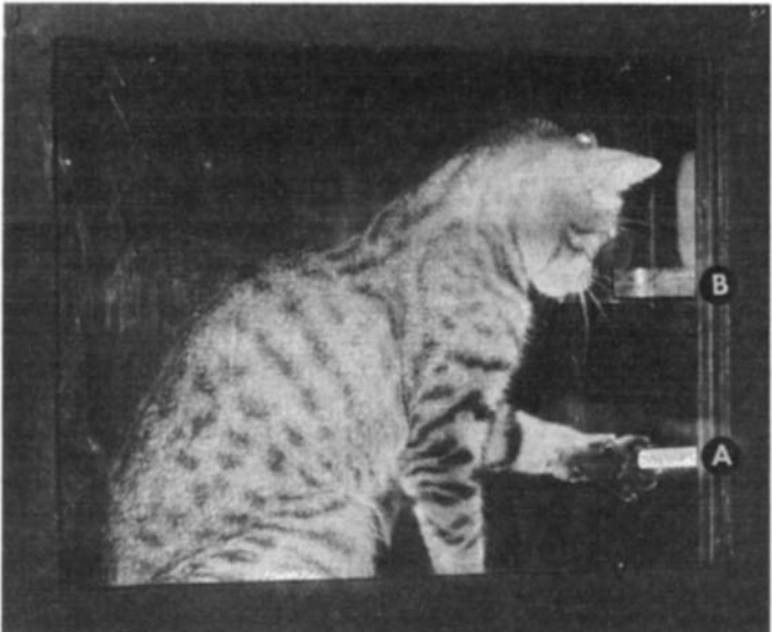
Researchers at the University of Delaware inhibited aggressiveness in cats by electrical stimulation of the caudate nucleus. (Plumer and Siegel 1973)
Electrical stimulation of the hypothalamus in the cats induced aggressive behavior toward rats in all 55 trials (100%).
Electrical stimulation of the caudate nucleus, applied 20 seconds before that, induced aggressive behavior toward rats in only 7 of 53 trials (11%).
The time it took to attack increased by 10-15 seconds, and the attacking time became much shorter as well.
When the cats received stimulation of the hypothalamus following that of the caudate nucleus, they circled the chamber and began the stalking phase of the attack.
But, instead of initiating a vicious biting attack toward the neck of the rat, the cats ignored the rat, often stepping on and over it without making any attempt to attack it.
Researchers at Bryn Mawr College inhibited aggressiveness in cats by electrical stimulation of the septal area. (Thomas and Evans 1983)
First, they stimulated the hypothalamus in the cats to induce emotional attack responses.
The cat would hiss, spit, have its fur stand up, and attack an object directly.
Electrical stimulation of the septal area had an extremely potent inhibitory effect on the emotional behavior.
When a manipulandum (a rod or a platform) that delivers electrical stimulation of the septal area was placed in the cage, the cats quickly learned to activate it to counteract the unpleasant hypothalamic effects.
Rats
An experiment using rats has shown that among the septal area, electrical stimulation of the lateral septum can inhibit aggression. (Brayley and Albert 1977)
In the experiment, the rats were first made aggressive by damaging their hypothalamus.
Next, the electrodes were implanted into the lateral septum, medial septum, and cingulate cortex in the rats.
Aggressiveness in the rats was then assessed 5 minutes before, during, and 5 minutes after the stimulation.
The test items were (a) presentation of a pencil just in front of the snout, (b) a sharp tap on the back with a pencil, (c) presentation of a hand before the snout, (d) gentle prods on the side of the body with a stick, (e) capture by the tail, and (f) grasping of the body.
Responses to these tests were quantified on a scale of 0–3.
They found that electrical stimulation of the lateral septum produced a dramatic decrease in the rats' aggressiveness by about 80%, while stimulation of the medial septum and cingulate cortex did not produce any noticeable changes.
Decrease in Aggressiveness
Monkeys
An experiment with monkeys has shown that electrical stimulation of the caudate nucleus reduced emotional components of pain. (Lineberry and Vierck 1975)
In the experiment, stump-tailed macaques were first restrained and trained to strike a panel with a force of at least 4.96 lbf (2.25 kg) when receiving an electric shock to their legs, thereby stopping the shock.
The force applied to the panel was then measured and considered an index reflecting the emotional aspect of pain.
The electric shock to the leg lasted for 15 seconds unless the monkey stopped it.
Electrical stimulation of the caudate nucleus in the stump-tailed macaques markedly reduced the force applied to the panel to escape the electric leg shock.
This was particularly pronounced in high-intensity electric shocks.
The inhibitory effect was only effective when caudate stimulation was 0-100 milliseconds ahead of the electric leg shock.
Applied Force
Disruption of Consciousness
Electrical stimulation of the claustrum has been shown to produce disruption of consciousness in humans and to produce a similar state in animals.
Humans
Norwegian neurophysiologist Carl Sem-Jacobsen repeatedly conducted human experiments of electrical brain stimulation on patients with schizophrenia and Parkinson's disease. (Sem-Jacobsen 1968)
Shem Jacobsen discovered that electrical brain stimulation can produce various levels of altered consciousness.
The regions that induced the responses are described as “between the Sylvian fissure and the pulvinar-posterior thalamic area,” which is rather extensive.
The regions also include the claustrum.
The changes observed in the patients' consciousness were divided into the following categories:
- Unconscious with or without grand mal seizure.
- Absence (brief loss of consciousness).
- Confused; chewing movements, smacking, swallowing; staring; on the border of losing consciousness.
- Reduced alertness; correct tracking capability limited to between 5 and 25 percent of the time.
- Drowsy; sleepy with or without yawning.
- Becomes alert; wakes up; regains full consciousness, activates thinking.
- Feeling of surroundings fading away; objects seem smaller; voice seems reduced. Drifting away toward fainting.
- Split personality; a feeling of split personality; has a sensation of being outside himself; split into two persons.
- Hallucinations; visual, auditory, or other forms of simple or complex hallucinations with or without other forms of psychotic behavior.
Below is an example of confusion caused by electrical stimulation in a male patient.
The patient, a member of the Norwegian Armed Forces in Canada during World War II, spoke some English, but for the past fifteen years he had lived on a small farm in a remote mountain valley.
During the electrical stimulation, he was asked to speak English for the records.
Stimulation of a particular electrode in this patient produced confusion. He could not give the investigator's name, but said in English, "I don't remember your name."
At the same time, he repeatedly moved his arms in a fashion he had been asked to do earlier, although he could not explain why he started moving his arms.
He was obviously confused. Yet, he remembered to speak English.
Drowsiness was induced in four patients.
The patients described it as "slightly more drowsy," "feels more tired," and "relaxes, gets drowsy."
Some patients slept during stimulation of certain electrodes without waking up. Stimulation of other electrodes, on the other hand, alerted the patients from sleeping without causing any unpleasant side effects.
Dr. Mohamad Koubeissi of the University of Washington reported a case in which electrical stimulation of the claustrum induced severe disruption of consciousness. (Koubeissi et al. 2014)
The patient was a 54-year-old woman with epilepsy who was undergoing electrical brain stimulation to locate the focus.
Stimulation of an electrode located between the left claustrum and insular cortex resulted in immediate impairment of consciousness, with arrest of reading, onset of blank staring, unresponsiveness to auditory or visual commands, and slowing of spontaneous respiratory movements.
This was repeated 10 times with 10 stimulation.
The patient returned to her normal state as soon as the stimulation stopped with no recollection of the events during the stimulation period.
Occasionally, the induced impairment of consciousness was associated with scanty, perseverative, and incomprehensible verbal output consisting of one or two syllables, with a confused look on the face.
Cats and Rats
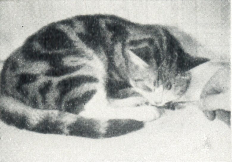
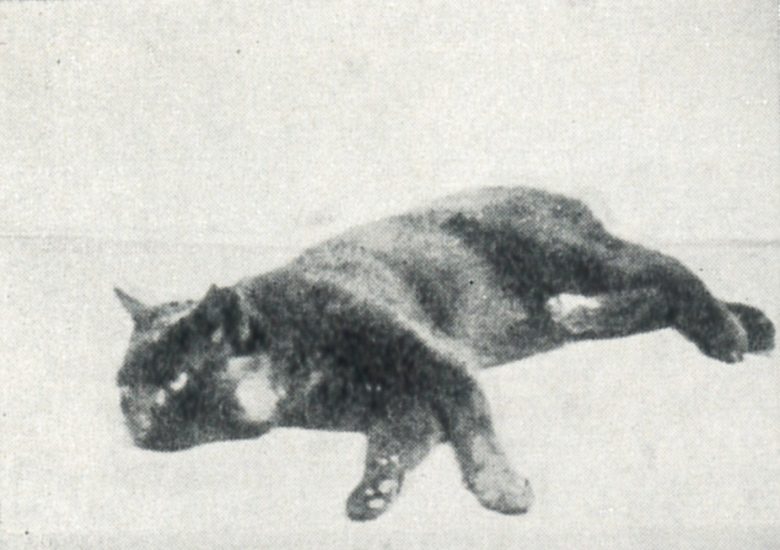
Researchers at Duke University induced an inactive state in cats by electrical stimulation of the claustrum. (Gabor and Peele 1964)
Prior to the stimulation, the cat reacted immediately and attentively to clicking noises and tapping on the cage.
15-20 seconds after the onset of the electrical stimulation of the claustrum, the cat became unresponsive to attempts at attracting its attention. This state continued beyond the period of stimulation.
Every time stimulation was applied, the cats performed the following stereotypical behavior:
First, the cat assumed a crouching position, flexing its legs under its body, then slightly turning its head to the opposite side occasionally. Finally, it would close its eyes and remain in this position for as long as 10 minutes after cessation of the stimulation, despite attempts at external distractions.
The cat was not asleep and its muscle tone was not reduced. It rather seemed unresponsive to moderate sensory stimuli and appeared to manifest an increased inertia with respect to movement.
The cat could be aroused from this state of inactivation only with loud noises and by shaking the enclosure. Once aroused in this manner, the cat returned to its pre-stimulation activities such as drinking milk.
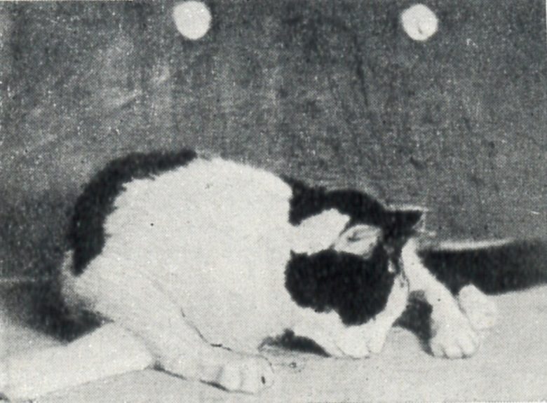
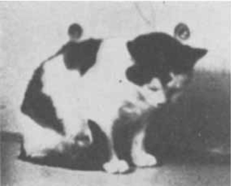
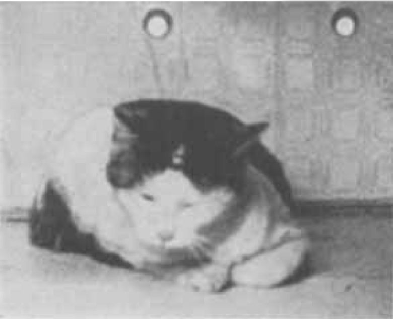
In an experiment with rats, Dr. Koubeissi confirmed that electrical stimulation of the claustrum can cause disruption of consciousness. (Bayat et al. 2018)
First, the rats learned that they could receive food pellets by responding to either a red or green light and sticking their noses into the correct port corresponding to the color.
When electrical stimulation of the claustrum in the rats was applied, their success rate in this task decreased, and the decrease was more pronounced as the stimulation intensity increased.
Disruption of Consciousness
As the stimulation intensity increased, the success rate decreased.
There were also 19 sessions in which about half of the rats had a 0% success rate.
This was due to immobility, apparent confusion, and stereotypic movements induced by the claustrum stimulation.
Some rats became inactive for up to 5 minutes and were unable to complete the session. The behavior of these rats resembled the disruption of consciousness observed in the patient following the claustrum stimulation.
The induced stereotypic movements were characterized by a sequence of movements: recoiled necks, hunched shoulders, flattening of the belly, and dragging of the body backward.
The rats then returned to their normal posture and fell into prolonged inactivity.
Motor Arrest
Electrical stimulation of the caudate nucleus has been shown to arrest ongoing movements. In human experiments, this sometimes caused confusion.
Humans
Dr. Van Buren's Study
Dr. John Van Buren of the National Institutes of Health conducted an experiment of electrical stimulation of the head of the caudate nucleus on patients with Parkinson's disease. (Van Buren 1963)
Electrical stimulation of the head of the caudate nucleus in the patients produced speech arrest and motor arrest, sometimes accompanied by confusion.
When the patients were stimulated while counting numbers or opening and closing their hands, those actions stopped.
When asked why, they usually gave trivial excuses unrelated to stimulation, leaving the impression that the impulse to speak or move had decreased.
In addition, some cases exhibited confused speech and behavior, along with memory loss related to them.
A 58-year-old male patient, Mr. E. B., stopped counting after receiving electrical stimulation of the caudate nucleus.
As an excuse he said, "I thought I had gone far enough," but it had been emphasized beforehand that he was to continue to count until told to stop.
Subsequently, there were observed abrupt arrests of speech with each stimulation. Although strictly reminded to continue counting, he continued to excuse his arrests variously, e.g., "It seemed like a barrier, a natural end."
In another session, he was asked to protrude his tongue and hold it, and continue slowly opening and closing his right hand. When given electrical stimulation, he withdrew his tongue and stopped the right-hand motion.
He later recalled stopping the right-hand motion but could not recall why he had done so.
Also in another session, he lay quietly for the first 3 seconds of stimulation, then abruptly sat up, turning slightly toward the examiner at his right.
At first he was unresponsive, fumbled with his shirt, and did not look at the examiner when spoken to.
Then, when asked why he had moved, he replied, "I wanted to sit up and talk to you. I wanted to tell you something."
Following a vague smile, he added, "Not very much, something less than nothing," and lay back upon the bed.
Within a minute he apparently had recovered fully but had no recollection of any part of the episode.
A 53-year-old female patient, Ms. H. T., also stopped counting after receiving electrical stimulation of the caudate nucleus, which was reproduced multiple times.
When asked why she had stopped counting she replied variously.
For example, "I thought you didn't want me to go on," "I thought I'd gone far far enough," and "I thought you were leaving the room."
In a session, when given stimulation, she turned her head to the left and made a humming noise in her throat. There was rapid blinking of the eyes.
Following the stimulation when asked how she felt, she replied, "One of the nurses was saying what they say in court, 'Y, Z, oh yea, oh yea.'"
No nurse had been present nor could speech from adjoining areas be heard.
A 58-year-old male patient, Mr. L.S., became confused after receiving electrical stimulation of the caudate nucleus.
He was lying quietly at the onset of stimulation and then suddenly turned his head and eyes to the right.
Following this he sat up and tried to remove the testing equipment from the left hand despite being told not to do so.
A few seconds later he became tractable to suggestion but would not respond verbally.
Shortly thereafter he recovered completely but had no memory of any part of the episode.
Tohoku University Study
Researchers at Tohoku University in Japan attempted to replicate the human arrest responses seen in Dr. Van Buren's study. (Kwak et al. 1978)
The subjects were pain patients and Parkinson's disease patients, and electrical stimulation of the head of the caudate nucleus was able to arrest speech and voluntary movements.
In rare cases, confusion also occurred.
The patients were asked to count or calculate and were given electrical stimulation while they performed the task.
The patients abruptly stopped counting immediately or within 1 or 2 seconds. In almost all cases immediately after the release of the stimulation the patient resumed counting as though no distraction had occurred.
For example, a patient was asked to count from one to ten.
When the patient reached the number three, electrical stimulation was given and the counting was interrupted until stimulation was released.
Following the release of stimulation the patient resumed counting from where he left off.
The patient was asked various questions while counting was suppressed by stimulation, and in almost all cases, after the release of the stimulation the patient was able to answer correctly the questions given during the stimulation
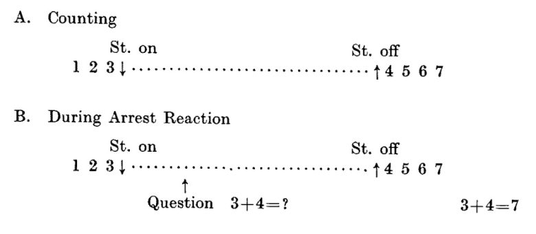
Furthermore, in almost all cases the patient could recall precisely all events occurring before, during and after the stimulation.
However, in three cases the patients could not recall all events occurring during the stimulation, and were not aware of stopping counting.
In such cases the patients did not resume counting after the release of the stimulation. Such confusion disappeared immediately or within 1 minute at most after the release of the stimulation.
After repeated stimulation the patients sometimes recounted from one or from a number that had already been counted, or mumbled something or occasionally arrest responses could not be induced.
The stimulation not only arrested speech but also voluntary movements.
For example, a patient was asked to lift his arms and to flex and extend his fingers repeatedly.
When electrical stimulation of the caudate nucleus was applied, motion of fingers was stopped but his arms remained lifted and shook lightly.
After the release of stimulation the patient could do voluntary movements as before the stimulation.
Cats
In animal experiments using cats, electrical stimulation of primarily the caudate nucleus has been observed to induce motor arrest responses.
The Montreal Neuroscience Institute found that electrical stimulation of the thalamus can arrest spontaneous movements in cats. (Hunter and Jasper 1949)
When stimulated while walking, the cat stopped as though frozen in place.

The cat remained in a fixed or crouched posture with a "staring" rather than "stunned" facial expression. The cat resumed walking as soon as the stimulation was withdrawn.
If eating, the cat suddenly stopped motionless, and if pursuing a mouse, the cat was also arrested in its pursuit.

There was no response to light or sound. The eyes remained open, often with slight twitching movements around the eyes and mouth.
Spinal reflexes and corneal reflexes remained unaltered.
Although there was paw withdrawal in response to pain during stimulation, the cats did not engage in defensive behavior, even in response to severe pain.
Researchers at the University of British Columbia, through electrical stimulation of the caudate nucleus, induced motor arrest responses in cats similar to those seen in the previous experiment. (McLennan et al. 1964)
Electrical stimulation of the caudate nucleus at low frequencies of less than 10 Hz stopped the cat's spontaneous movement.
The cat was immediately frozen in the middle of a movement at the onset of stimulation even to the extent of holding one leg flexed in the act of making a step.
A continued twitching of the tail was, however, frequently noted.
If the cat was advancing toward a piece of food at the beginning of the stimulation period its attention was not diverted and it continued to gaze fixedly upon its goal.
As soon as the stimulation was ended the animal moved forward without hesitation.
Increasing the frequency of stimulation gave rise in all animals to rotational movements.
At frequencies of about 10-15 Hz, the cat raised its head and turned the head and neck, as if it looked over the shoulder opposite to the side of stimulation.
With a further increase in frequency the cat began to circle in the opposite direction, the speed of circling being frequency-dependent.
Electrical stimulation of the caudate nucleus has also been shown to inhibit learned avoidance behavior. (STEVENS 1961)
In the experiment, the cats were first trained to pass through a small open doorway to avoid electric shock applied a few seconds after a buzzer sounded.
Next, when electrical stimulation was applied to the caudate nucleus 20 seconds before the buzzer sounded, the cats no longer took the avoidance behavior they had learned.
When the buzzer sounded, the cat sometimes raised a forepaw as though about to go through the doorway, but then stopped, looked around, and failed to cross.
Electrical stimulation of the caudate nucleus has also been shown to inhibit food-retrieving movements. (Buchwald et al. 1961)
In the experiment, the cats first learned that pressing a lever would reward them with a small amount of liquid cream.
Electrical stimulation of the caudate stimulation at low frequency of 0.2 Hz produced first a slowing and then a cessation of the cat's lever-pressing response.
Increasing the stimulation frequency up to 5 Hz resulted in increasing the effect. In some cases, the stimulation caused an abrupt cessation of lever-pressing.
Following cessation of lever-pressing the cat appeared to be unconcerned with the lever, although licking of cream remaining in the food cup would often continue.
At the termination of the low-frequency stimulation, the cat usually remained in a sitting position for a few moments and then walked over and began to push the lever.
When high-frequency stimulation of 300 Hz was delivered to a cat that had stopped lever pressing due to low-frequency stimulation, an immediate return to lever pressing occurred.
That is, the cat's lever pressing could be turned off by low-frequency stimulation and turned on by high-frequency stimulation.
Other Animals
In experiments using other animals, such as monkeys, it has also been observed that electrical stimulation of primarily the caudate nucleus can induce motor arrest responses.
Researchers at the University of Rochester arrested monkey movements by electrical stimulation of the anterior cingulate cortex. (Smith 1945)
When electrical stimulation of the anterior cingulate cortex was applied to the rhesus monkeys, any ongoing movement immediately ceased, and their muscle tension relaxed.
Even a monkey struggling after drug administration could be completely sedated by stimulating the anterior cingulate cortex.
Following stimulation all movements ceased, the eyes closed, and the monkey remained in a quiescent state resembling sleep for some minutes.
Researchers at the University of Paris arrested monkey movements by electrical stimulation of the caudate nucleus. (Kitsikis 1968)
While seated in a restraint chair, the monkeys performed a variety of arm movements, as shown in the diagram below.
All these movements were performed to obtain a piece of grape.

Electrical stimulation of the head of the caudate nucleus was able to arrest all these movements.
The more effortful the movement, the easier it was to stop it.
That is, movements such as reaching a high distance or pulling a weight could be stopped with weaker stimulation.
In addition, preventing the initiation of a movement required weaker stimulation than stopping the movement itself.
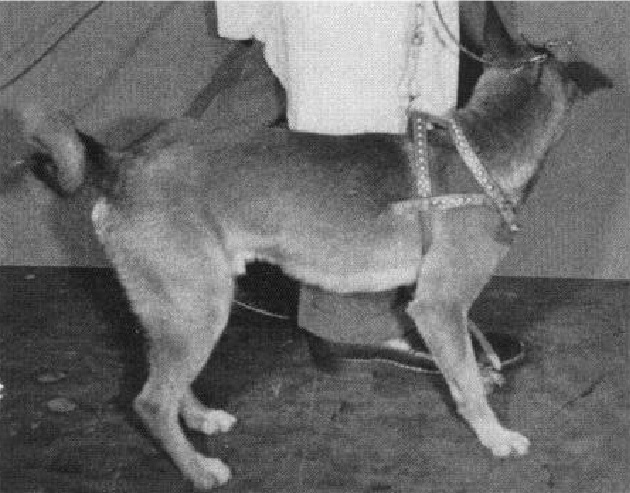
An experiment with dogs has also shown that electrical stimulation of the caudate nucleus can induce motor arrest responses. (Goldzband et al. 1951)
Electrical stimulation of the caudate nucleus arrested spontaneous walking in the dogs.
With the onset of the stimulation the dog simply completed the step he was making and then halted.
Usually, but not always, other patterned movements such as tail wagging were also inhibited.
The walking arrest was sometimes accompanied by several simple movements, such as head turning and rotational movements.
For example, stimulation of one electrode consistently resulted in walking arrest accompanied by head turning to the left.
In an experiment with rabbits, electrical stimulation of the caudate nucleus has been observed to produce general behavioral inhibition. (Klemm and Dreyfus 1975)
Electrical stimulation of the caudate nucleus produced the following inhibitory effects on behavior in the rabbits.
- They jiggled less when confined in a box.
- They moved around less when allowed to do so freely.
- They took longer to get up when being turned over.
- They no longer avoided an air blast, which is an aversive stimulus.
Amnesia
Electrical stimulation of the hippocampus, amygdala, and caudate nucleus has been shown to induce amnesia in rodents.
Hippocampus, Amygdala
Electroconvulsive shock, in which electric current is passed through the brain via electrodes placed on the scalp, causing a generalized seizure, is known to cause amnesia. (Kopp et al. 1966)
Experiments on rodents have shown that similar effects can be induced by electrical stimulation of specific sites of the brain.
A Columbia University study found that electrical stimulation of the hippocampus in mice can induce amnesia similar to that caused by electroconvulsive shock. (Lidsky and Slotnick 1970)
First, a metal bar was mounted on one wall of the cage that would deliver an electric shock if touched.
The mice were allowed to explore the cage and learn that touching the bar would result in an electric shock.
The mice then received electrical stimulation of the hippocampus or the neocortex area, or electroconvulsive shock.
The following day, when the mice were allowed to explore the cage again, those that had received the hippocampal stimulation took on an appearance of amnesia similar to those that had received electroconvulsive shock.
That is, the mice seemed to have forgotten that touching the metal bar would result in an electric shock.
This was indicated by a decrease in the time taken to contact the bar, an increase in exploratory behavior (increased "vertical movement" and "activity" below), and a decrease in the number of freezing.
Amnesia
Electrical stimulation of the amygdala has also been shown to cause amnesia in rats. (Bresnahan and Routtenberg 1972)
First, they electrified the cage floor and placed a platform on top of it so that if the rats stepped off it they would receive an electric shock.
When the rats were placed on this platform, they quickly learned that stepping down on the floor would result in an electric shock.
When the rat returned to the platform, electrical stimulation of the amygdala was then applied.
The following day, when the rats were placed on the platform again, those that had received electrical stimulation to the amygdala were considerably faster to step down on the floor than those that had not.
The rats seemed to have forgotten that stepping down on the floor would result in an electric shock, suggesting that amnesia had occurred.
In addition, within the amygdala, stimulation of the medial nucleus was most effective.
Amnesia
The amygdala stimulation considerably shortened the time taken to descend to the floor after receiving an electric shock.
Caudate Nucleus
A research group at Stony Brook University found that electrical stimulation of the caudate nucleus in rats causes amnesia. (Wyers et al. 1968)
A lever was placed in the cage, and the rats could press it to receive sweet water.
Upon the 50th lever press, the rats received an electric foot shock.
After a delay of 0.1, 1, 5, or 30 seconds, electrical stimulation of the caudate nucleus was applied.
The next day, the rats were placed back in the cage, and the rats who had received caudate stimulation pressed the lever to take the sweet water with almost no hesitation.
The time taken to drink was almost the same as that measured before the shock.
In contrast, the rats who had not received caudate stimulation were very vigilant toward the lever and showed a marked delay in taking the sweet water.
This amnesia induced by the caudate nucleus did not differ largely depending on the delay time between the electric shock and brain stimulation.
Amnesia
The caudate stimulation markedly shortened the time taken to drink, which did not differ largely depending on the delay time between the electric shock and brain stimulation.
In the subsequent experiment, the research group showed that amnesia caused by caudate nucleus stimulation depended on the delay between the electric shock and the brain stimulation. (Wyers and Deadwyler 1971)
This was achieved by making the delay time longer than in the previous experiment, from the maximum of 30 seconds to 300 seconds.
In the experiment, the rats were allowed to lick the water from a water dispenser mounted on the cage, and upon the 50th lick, the rats received an electric foot shock.
After that, electrical stimulation of the caudate nucleus was applied after a longer delay of 30, 120, or 300 seconds.
The following day, when the rats were placed back in the cage, the rats that had received caudate stimulation licked the water with almost no hesitation, confirming the results of the previous experiment.
The difference from the previous one was that the shorter the delay between the electric shock and the brain stimulation, the more severe the amnesia.
Amnesia
The caudate stimulation markedly shortened the time taken to drink, and this was more effective the shorter the delay time between the electric shock and brain stimulation.
Electrical stimulation of the caudate nucleus has also been shown to impair learning in rats. (Peeke and Herz 1971)
In the experiment, the rats ran through a Lashley III maze, aiming for wet mash placed at the goal.
Success was made by not taking a wrong turn at any branch point during a single trial, and the rats underwent maze learning until they achieved four successes out of five trials.
During maze learning, the rats received single or multiple electrical stimulations of the caudate nucleus, either after reaching the goal or after crossing each branch point.
They found that this impaired the maze learning in the rats and increased the number of trials taken to reach the criterion, especially when multiple electrical stimulations were given at once after reaching the goal.
Amnesia
The caudate stimulation impaired the maze learning in rats and increased the number of trials taken to reach the criterion.
Sleep, Adynamia
Electrical stimulation of the caudate nucleus, thalamus, and basal forebrain has been shown to induce sleep.
Electrical stimulation of the anterior hypothalamus and caudate nucleus has also been shown to induce an inactive state accompanied by loss of muscle tone, called adynamia.
Humans
Caudate Nucleus
Psychiatrist Robert Heath of Tulane University found, in an experiment on a rhesus monkey and a schizophrenic patient, that electrical stimulation of the head of the caudate nucleus can induce sleep. (Heath and Hodes 2017)
In the rhesus monkey, when weak electrical stimulation at 10 Hz was applied to the head of the caudate nucleus, the monkey became drowsy and soon went into deep sleep.
The brainwave patterns were not that different from those seen during normal sleep.
In the schizophrenic patient, electrical stimulation of the head of the caudate nucleus at 100 Hz for two minutes resulted in drowsiness, which progressed rapidly into deep sleep.
It was impossible to arouse the patient.
Clapping of hands near the patient's ear elicited only minimal reaction. This persisted for a few minutes after cessation of the stimulation.
A gradual awakening then began and in the course of 8-10 minutes, the patient appeared to be at about the level of alertness he exhibited prior to stimulation.
Subthalamic Nucleus
Soviet scientist Natalia Bekhtereva showed, in an experiment on patients with movement disorders, electrical stimulation of the subthalamic nucleus can induce drowsiness. (Bekhtereva 1969)
In one patient, repetition of low-frequency (4, 10 Hz) and high-frequency (25, 50 Hz) electrical stimulation of the subthalamic nucleus with 15-30 second intervals was administered.
As a result, wakefulness and drowsiness alternated.
The patient, who had just been lively, became limp, yawned, complained of drowsiness, began to doze, and then, without any intervention from the examiner, became lively and alert again.
In another patient, four high-frequency (50 Hz) electrical stimulations to the subthalamic nucleus with 15-30 second intervals produced brief drowsiness.
Cats
Thalamus
Dr. Walter Hess of the University of Zurich in Switzerland discovered that electrical stimulation of the thalamus and hypothalamus in cats can induce sleep. (Hess 1958)
When electrical stimulation of the interthalamic adhesion at a low frequency of 4-6 Hz was delivered, the cat would settle down to sleep in a typical attitude.
The cat would often open its eyes when the cat was struck or shaken.
If meat was placed before its nose, the cat would very quickly prepare to seize it. After the cat ate the food, it would soon lie down again and go back to sleep.
Upon the stimulation the cat did not appear to "pass out" suddenly, but would seek a comfortable position, which was sometimes not found until after a few postural adjustments.
Anterior Hypothalamus
Electrical stimulation of the anterior hypothalamus induced an inactive state distinct from sleep, which Dr. Hess termed adynamia. (Hess 1958)
Upon the stimulation there was a lowering of blood pressure, and the cat would sink from the posture being maintained at the time to assume an extraordinary position.
The tone of the skeletal muscles, as well as spontaneous activity and responsiveness, was entirely lost. When touched by the experimenter, the cat would remain entirely unreactive.

Caudate Nucleus
Other researchers, also of the University of Zurich, showed that electrical stimulation of the head of the caudate nucleus can also induce adynamia. (AKERT and ANDERSSON 1951)
Within seconds to 2-3 minutes after the caudate stimulation, the previously lively cat would become increasingly calm, sit down or lie down, and constantly squint. Spontaneous activity appeared extinguished.
It would only react to external stimuli by turning its head. Otherwise, the cat would display a tired, disgruntled expression, which contrasted strikingly with its active head posture.
Based on its overall appearance, one would expect the cat to lie down completely and curl up to sleep at any moment.
In fact, however, the cat would remain motionless in a sitting or lying position with its head raised, remaining receptive to all sensory stimuli. This continued for minutes after the stimulation had ceased.
Basal Forebrain Synchronizing Area
A UCLA study found that electrical stimulation of the basal forebrain synchronizing area produces a powerful sleep-inducing effect in cats. (Sterman and Clemente 1962, Sterman and Clemente 1962)
The basal forebrain synchronizing area is a region where electrical stimulation can induce brainwave synchronous activity in the cerebral cortex, similar to that observed during sleep.
Specifically, this was the region extending from the diagonal band of Broca to the lateral preoptic area.
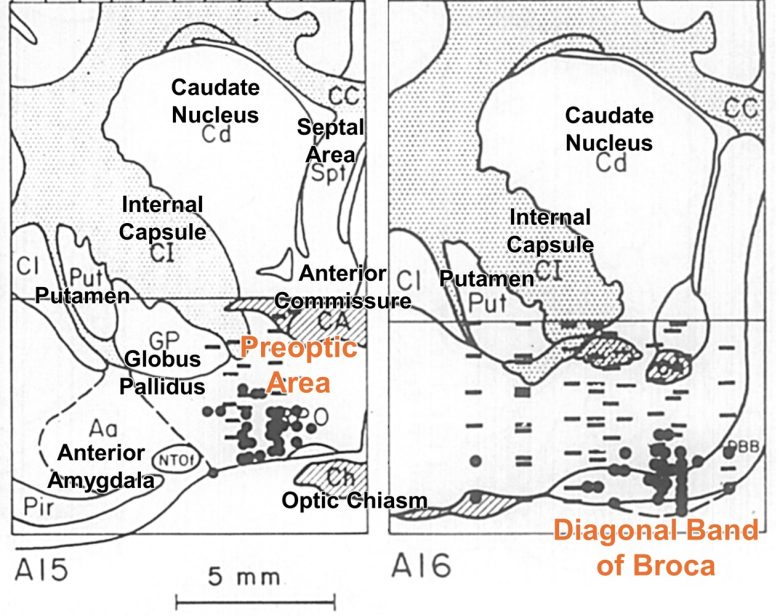
The region lay somewhat ahead of the anterior hypothamamic region where Dr. Hess had induced adynamia by electrical stimulation.
Stimulation delivered to the basal forebrain synchronizing area resulted in a surprisingly rapid transition from wakefulness to drowsiness and sleep.
The sequence of events initiated by the onset of stimulation usually consisted of an arrest of ongoing behavior, followed by retreat to some corner of the cage, and the assumption there of a natural reclining position.
This was followed by an apparent loss of interest in the immediate environment.
If undisturbed, the reclining cat drooped his head to the floor or rested it on his forepaws, closed or partially closed his eyes, and, to all appearances, went to sleep.
The time from the onset of stimulation to the occurrence of sleep ranged from as short as 5 seconds to as long as 3 minutes, with an average duration of approximately 30 seconds.
If the cats were left undisturbed, they continued sleeping.
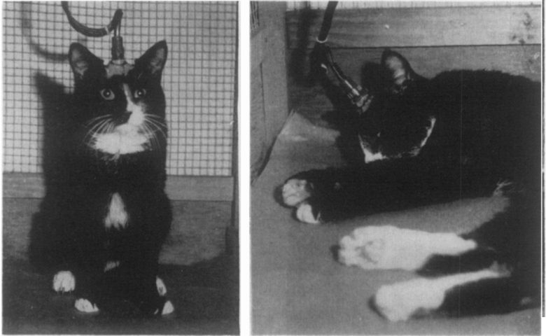
Furthermore, electrical stimulation of the basal forebrain synchronizing area was powerful enough to stop a cat from attacking a rat and put it to sleep.
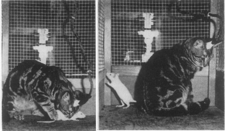
A study at the University of Delaware confirmed that electrical stimulation of the basal forebrain synchronizing area can induce sleep. (Lineberry and Siegel 1971)
In the experiment, the cats were first fasted for 24 hours and then given food.
When basal forebrain stimulation was initiated a few seconds after the cat began eating, it would stop eating, walk away from the food dish, and lie down.
If the stimulation was continued for 45-50 seconds, the cat would go into sleep and continue to sleep after the stimulation was discontinued.
Monkeys
Experiments with monkeys have also shown that electrical stimulation of the anterior hypothalamus can induce adynamia (an inactive state accompanied by loss of muscle tone).
Hypothalamus
A study at the University of Colorado showed that electrical stimulation of the anterior hypothalamus can produce adynamia in rhesus monkeys. (Sheer 1961)
With anterior hypothalamic stimulation, the monkey became distinctly quiescent.
In a free-moving situation he was inactive; left to himself, he generally sat quietly and sometimes closed his eyes.
In a restraining cage, he usually closed his eyes and appeared to doze. He could be activated by various stimuli, but his reactions were less intense and generally of shorter duration than normal.
The salient features of his behavior during the stimulation could be characterized as lowered activity, decreased reactivity to stimuli, and general quiescence.
The behavior was accompanied by pupillary constriction and a generalized loss of muscular tone, resembling the state described by Dr. Hess as adynamia.

Jose Delgado of Yale University also showed that electrical stimulation of the anterior hypothalamus can produce adynamia in rhesus monkeys. (Delgado 1964)
The stimulated monkey showed quiescence and dozing, but there was also a generalized loss of muscular tone, and this effect is similar to the adynamia described by Dr. Hess rather than natural sleep.

Food Refusal
Electrical stimulation of the caudate nucleus and hypothalamus has been confirmed to induce food refusal.
Delgado's Animal Experiments
Rhesus Monkeys
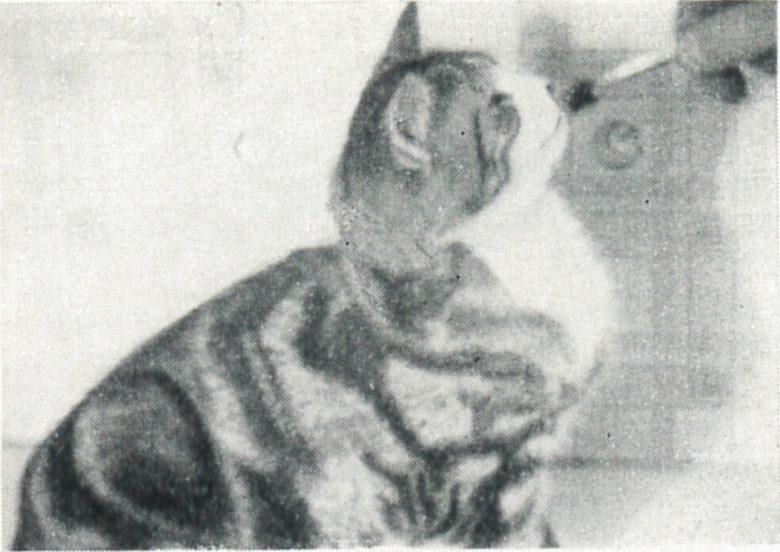
Delgado showed that electrical stimulation of the head of the caudate nucleus can inhibit appetite in monkeys. (Rubinstein and Delgado 1963, Delgado 1964)
When the rhesus monkeys were restrained in a chair and the caudate nucleus was electrically stimulated, the behavior of approaching, taking up, and eating food was inhibited.
When fruit was offered, the monkey turned its head and looked at the food without making any effort to take it, even when very close.
The inhibitory effect was observed with all kinds of food tested, which included peanuts, pellets, apples, bananas, and oranges, and attempts by the experimenters to force the monkey to eat failed, with the monkey turning away from the food.
Biting or chewing also stopped as soon as the caudate stimulation started, but was resumed as soon as the stimulation ceased.
If the monkey was walking toward the food, it would hesitate, stop, and slowly move away when stimulated.
If the monkey already had the banana in its hand or mouth, the fruit was dropped to the floor of the cage as soon as the stimulation started, and the monkey showed no further interest in it, neither threatening nor retaliating when the other monkeys took the banana.
The monkey spit out any food in the mouth, and if the monkey had its pouches filled with food, it tried actively to empty it with the paws.
If the monkey was drinking, it stopped as soon as the stimulation started, slowly averted its head, and moved away from the drinking tube.


Gibbons
In an experiment with gibbons as well, Delgado showed that electrical stimulation of the caudate nucleus can inhibit appetite. (Delgado 1975)
The voracious eating of an apple stopped as soon as the head of the caudate nucleus was stimulated and the piece of fruit was slowly withdrawn from the mouth.
The animal remained peaceful for the duration of stimulation, resuming eating a few seconds after its cessation.
In addition, lever pressing to obtain food pellets was also inhibited by caudate nucleus stimulation.


Other Animal Experiments
Caudate Nucleus
A study at the University of Delaware showed that electrical stimulation of the caudate nucleus can inhibit appetite in cats. (Lineberry and Siegel 1971)
In the experiment, the cats were first fasted for 24 hours and then given food.
Upon the stimulating of the caudate nucleus, the cat stopped eating and stood quietly with its head poised over the food dish.
The cat was not frozen but was able to look around and locomote.
Medial Hypothalamus
Researchers at the University of Pennsylvania showed that, in an experiment with rats, electrical stimulation of the medial hypothalamus and lateral hypothalamus had opposing effects on feeding. (Smith 1961)
Electrical stimulation of the medial hypothalamus caused inhibition of food intake, even when the rats were highly deprived.
This inhibitory effect was particularly pronounced during the first 30 minutes when food was given after food deprivation.
In contrast, electrical stimulation of the lateral hypothalamus induced a facilitation of food intake, resulting in eating or chewing that lacked the quality of discrimination.
The rats seized, put into their mouths, and chewed anything they happened to encounter, whether it was food, wood chips, feces, or paper clips.
They then dropped the objects and seized something else until the end of stimulation, when the behavior stopped.
Researchers at the University of Tennessee made similar observations in an experiment with rats. (Morgane 1961)
Electrical stimulation of the medial hypothalamus inhibited food intake, and even the hungry rats stopped eating when stimulated.
In contrast, electrical stimulation of the lateral hypothalamus facilitated food intake, and even satiated rats began eating when stimulated.
Simultaneous stimulation of the lateral hypothalamus, which facilitates appetite, and the medial hypothalamus, which inhibits appetite, cancelled the effect of each other out.