Fear

Electrical stimulation of specific sites of the brain can cause fear and flight responses in humans and animals.
The well-known sites are the amygdala, hypothalamus, and periaqueductal gray (PAG) of the midbrain, the same sites that elicit rage and aggressive behavior, but in detail there is a topographical separation. Fear, however, is widely induced from other sites as well.
In addition, more dangerous symptoms such as panic attacks and suicidal thoughts may also occur.
Panic attacks are sudden, unpredictable episodes of intense fear and are known to correlate with suicidal thoughts and attempts. (Weissman et al. 1989)
If abused, this would even make possible an extremely heinous criminal act where a homicide is made out to be a suicide without leaving any evidence.
Table of ContentsAll_Pages
Circuitry
Multiple Fear Circuits
The fear circuit runs from the amygdala through the hypothalamus to the periaqueductal gray (PAG) of the midbrain. (Panksepp 1998)


Three major brain sites related to fear. The amygdala, hypothalamus, and PAG.
(Modified from Panksepp 1998, the brain image from Vanderah 2018)
Electrical stimulation of these three regions has been confirmed to produce fear responses in both humans and animals.
In particular, electrical stimulation of the hypothalamus and PAG has been shown to produce intense fear, such as panic attacks.
It is known that fear can be subdivided into several types, each of which consists of a different neural circuit. (Gross and Canteras 2012)
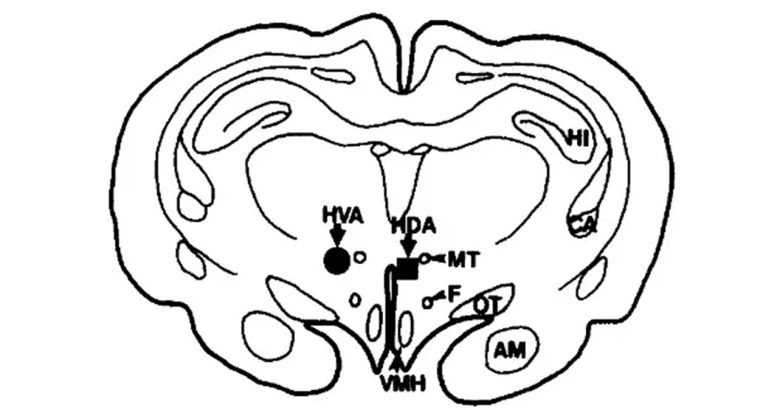
For example, the neural circuit for fear of predators, that is, innate fear, runs from the medial nucleus of the amygdala through the ventromedial nucleus of the hypothalamus to the dorsal part of the PAG in the midbrain.
On the other hand, the neural circuit for fear of stimuli associated with pain, that is, learned fear, runs from the lateral nucleus and basal (basolateral) nucleus through the central nucleus of the amygdala directly to the PAG.
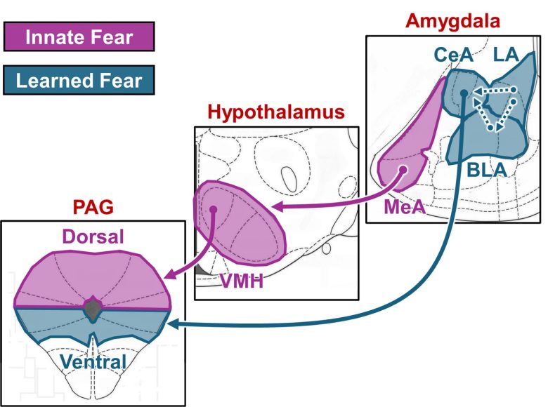
Neural circuits for innate fear and learned fear.
(Modified from Gross and Canteras 2012, the brain atlas from Paxinos and Watson 2009)
MeA = medial nucleus of the amygdala; LA = lateral nucleus of the amygdala; BLA = basal (basolateral) nucleus of the amygdala; CeA = central nucleus of the amygdala; VMH = ventromedial nucleus of the hypothalamus; PAG = periaqueductal gray.
Animal experiments have shown that electrical stimulation of both the medial and lateral regions of the amygdala can produce fear responses, and as I explained, this is thought to be the result of activating different types of fear circuits.
Separation of Fear and Rage
The neural circuit for innate fear is similar to that for defensive rage (rivalry aggression), but a closer look reveals a topographical separation.
In the medial amygdala, the posteroventral part is involved in innate fear, while the posterodorsal part is involved in defensive rage. (Gross and Canteras 2012, Raam and Hong 2021)
In the ventromedial nucleus of the hypothalamus, the dorsomedial part is involved in innate fear, while the ventrolateral part is involved in defensive rage. (Gross and Canteras 2012, Hashikawa et al. 2017)
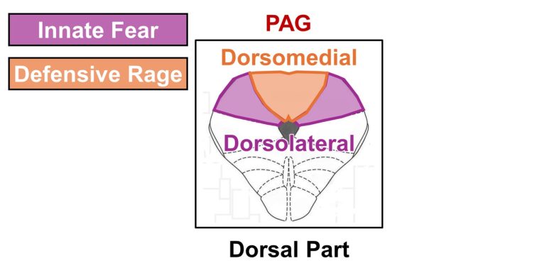
In the dorsal part of the PAG, the dorsolateral part is involved in innate fear, while the dorsomedial part is involved in defensive rage. (Gross and Canteras 2012, Li et al. 2025)

Separation of the neural circuits for fear and rage
(The brain atlas from Paxinos and Watson 2009)
MeA = medial nucleus of the amygdala; VMH = ventromedial nucleus of the hypothalamus; PAG = periaqueductal gray.
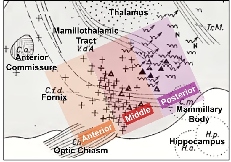
Three Regions in the Hypothalamus
The hypothalamus can be divided into three regions: the anterior, middle, and posterior regions.
The middle region contains the above-mentioned ventromedial nucleus, which is involved in the innate fear (fear of predators). (Gross and Canteras 2012)
In addition, the anterior region contains the anterior hypothalamic nucleus, and the posterior region contains the dorsal premammillary nucleus, both of which are also involved in innate fear.
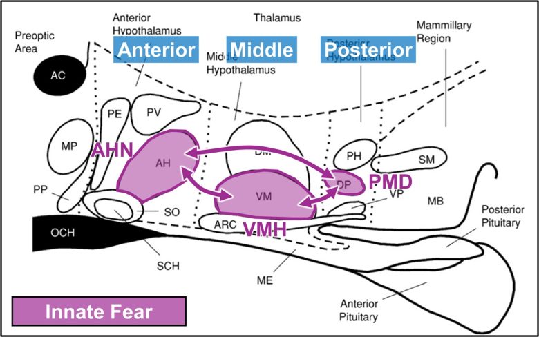
The regions that are involved in innate fear in the hypothalamus
(Modified from Halász 2004)
AHN = anterior hypothalamic nucleus; VMH = ventromedial nucleus of the hypothalamus; PMD = dorsal premammillary nucleus.
Electrical stimulation of any regions of the anterior, middle, or posterior hypothalamus has been shown to induce panic attacks or panic attack-like intense escape responses.
Hippocampus
The hippocampus is well known for its involvement in memory, but it is also involved in emotions. (Strange et al. 2014)
The dorsal part of the hippocampus mediates cognitive functions, particularly spatial memory, whereas the ventral part of the hippocampus mediates emotional responses.
It is thought that the ventral hippocampus, through its connections with the hypothalamus, can generate innate fear independently of the amygdala.
This connection is via the lateral part of the septal area, and the ventral hippocampus supplies nerves to a wide region of the lateral septum.
The lateral septum has been found to be capable of both suppressing and promoting fear responses, depending on subregion differences. (Rizzi-Wise and Wang 2021)
There are separate pathways from the hippocampus that connect to the two subregions.
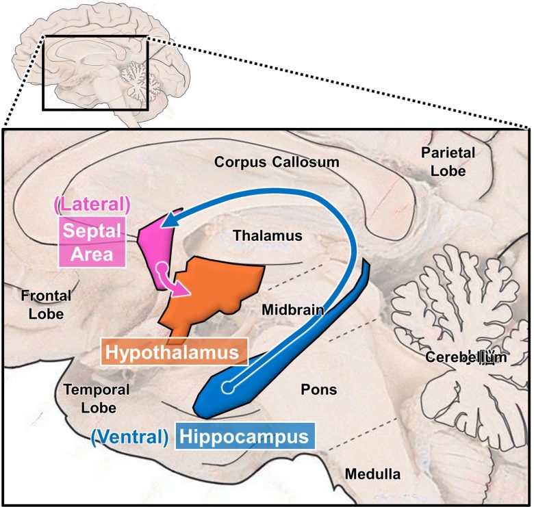

The ventral hippocampus connects to the hypothalamus via the lateral septum and influences emotions.
(Modified from Strange et al. 2014, the brain image from Vanderah 2018)
Electrical stimulation of the hippocampus has been confirmed to induce fear responses in both humans and animals.
Other Regions
Other than that, it has been observed that electrical stimulation of the nucleus accumbens and anterior cingulate cortex induces fear responses in humans.
The anterior cingulate cortex is thought to be involved in modulating fear expression through activation of the amygdala. (Milad et al. 2007)
The nucleus accumbens has been shown to generate opposing emotions of desire and fear from different regions in the anterior-posterior direction. (Berridge and Kringelbach 2015)
It has also been confirmed that electrical stimulation of the substantia nigra and subthalamic nucleus can induce depression.
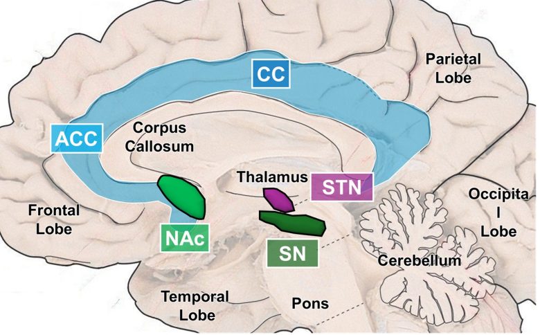
Other regions that induce fear and depression
(The brain image from Vanderah 2018)
CC = cingulate cortex; ACC = anterior cingulate cortex; NAc = nucleus accumbens; SN = substantia nigra; STN = subthalamic nucleus.
Humans
Electrical stimulation of the amygdala, hypothalamus, and periaqueductal gray (PAG) of the midbrain has been shown to induce a sense of fear in humans.
More dangerous symptoms can also occur, such as suicidal thoughts and panic attacks.
In addition, it has also been shown that fear was obtained from the hippocampus, anterior cingulate cortex, and nucleus accumbens, and depression was obtained from the substantia nigra and subthalamic nucleus.
Panic Attacks (Sudden Intense Fear)
PAG
At Duke University Medical Center, destruction of the midbrain tegmentum was applied to treat patients with intractable pain. (Nashold et al. 1969)
Prior to the operation, an experiment of electrical stimulation of the midbrain tegmentum was conducted.
The stimulation primarily produced a sense of pain, but also a sense of fear from specific sites.
Specifically, when electrical stimulation was applied to the PAG in the midbrain tegmentum, the patients experienced intense fear.
This sensation was extremely unpleasant, and although the patients were able to tolerate the intense pain caused by the stimulation, they were unable to tolerate this sense of fear.
They described it as "fearful," "frightful," or "terrible," and they would become apprehensive and not allow further stimulation.
At Tokyo Women's Medical University in Japan, an experiment was conducted on pain patients to investigate the analgesic effect of electrical stimulation of the PAG. (Amano et al. 1982)
Two types of electrical stimulation were used: low-frequency stimulation of 1-20 Hz and high-frequency stimulation of 50 Hz, but unfortunately neither had any analgesic effect.
However, high-frequency stimulation produced dramatic changes in the patients' moods.
The patients suddenly expressed a very strong sensation of fear and a burning, hot feeling of the entire body.
All of them appeared to be in a confused and acutely perplexed state.
The patients described themselves, "Something horrible is coming," "Somebody is now chasing me, I am trying to escape from him."
Another patient stated that he had an abrupt feeling of uncertainty "just like entering into a long, dark tunnel."
In addition, pulse rates and blood pressure increased significantly, pupil dilation and facial flushing occurred, and all the subjects experienced nystagmus.
When the stimulation was off the PAG and in the reticular formation, they felt a strong pain in their faces, trunks, and limbs on the opposite side.
Hypothalamus
At the Carlo Besta Neurological Institute in Milan, electrical stimulation of the posterior hypothalamus was administered to eight patients with cluster headaches. (Franzini et al. 2004)
All the patients felt a strong sense of fear, making statements such as, "“I feel near to death," and "I am at the edge of the end."
Continuous application of reduced intensity stimulation was successful in completely eliminating the pain of cluster headaches.
Following this report, at the University of Liège, Belgium, electrical stimulation of the posterior hypothalamus was also administered to six patients with cluster headaches. (Schoenen 2005)
During the test stimulation for one patient, a panic sensation with rapid breathing, tachycardia and moderate hypertension occurred.
Another patient fell into a deep coma due to a massive brain hemorrhage a few hours after surgery and died three days later.
One patient experienced only temporary relief, but the remaining three patients showed favorable progress.
At four German medical centers, electrical stimulation of the posterior hypothalamus was also administered to six patients with cluster headaches. (Bartsch et al. 2008)
In two patients, panic attacks occurred while the electrical stimulation parameters were adjusted.
With the application of continuous electrical stimulation, three patients became attack-free of cluster headaches, while three patients failed treatment.
Two cases were reported at a German university hospital in which treatment of cluster headache with electrical stimulation of the posterior hypothalamus failed. (Pinsker et al. 2008)
In test stimulation in both patients, high-intensity electrical stimulation of the posterior hypothalamus produced panic attacks accompanied by autonomic disturbance.
Although pain relief was observed for a few days after starting continuous stimulation, the follow-ups several months later showed no significant reduction in intensity or frequency of headaches.
Researchers at West Virginia University conducted an experiment on electrical stimulation of the ventromedial nucleus of the hypothalamus (VMH) in order to explore the neural circuit for panic attacks. (Wilent et al. 2010)
The VMH is located just anterior to the posterior hypothalamic region that has been shown to induce panic attacks in the previous studies.
The subject was a 50-year-old woman scheduled to have stimulation electrodes implanted in the hypothalamus for obesity treatment.
Electrical stimulation of the VMH induced a response that met the criteria for a DSM-IV panic attack.
Within 5–20 seconds of the onset of stimulation, a dramatic increase in anxiety was obvious and the patient reported troubled feelings to this effect, e.g., saying, “I am getting very emotional.”
This was accompanied by hyperventilation, shortness of breath, an increase in blood pressure, an increase in heart rate, and some nausea.
All symptoms subsided within 10–90 seconds of stimulation cessation.
As the electrode progressed deeper into the VMH, the intensity of stimulation required to induce an attack episode decreased.
Furthermore, stimulation that appeared to be in the center of the VMH was more potent.
Nucleus Accumbens
A research group at the University of Florida reported a case in which electrical stimulation of the nucleus accumbens produced a sensation of fear and panic. (Shapira 2005)
The patient was a 52-year-old man with obsessive-compulsive disorder, and he experienced reproducible sensations of flushing, fear, and "panic" with electrical stimulation of the right nucleus accumbens.
He described the sensation as feeling hot, flushed, fearful, and "panicky."
He could feel palpitations in his chest, and when asked indicated he had an impending sense of doom.
This sensation began when stimulation began and rapidly dissipated when it stopped.
The sensation of panic was accompanied by tachycardia: heart rate increased rapidly from 53 to 119 beats per minute upon initiation of stimulation and returned to 74 beats per minute after stimulation was discontinued.
The research group studied the responses induced by electrical stimulation of the nucleus accumbens and anterior limb of the internal capsule in five patients with obsessive-compulsive disorder. (Okun et al. 2006)
Mainly from the ventral part, both non-mood-related and mood-related responses were induced.
Non-mood-related responses included taste, smell, and smile.
Mood-related responses included autonomic changes, increased breathing rate, sweating, nausea, cold sensation, heat sensation, and fear and panic attacks.
Fear and panic responses appeared clustered around the most ventral electrode. Two of the patients who received this stimulation experienced severe panic attacks.
Higher stimulation intensity was associated with increased hallucinations, e.g., smell and taste, worsened mood, and increased anxiety.
Suicides
Hypothalamus
At Massachusetts General Hospital, electrical stimulation of the posterior hypothalamus was administered to a 43-year-old female patient with chronic migraine as an experimental treatment. (Walcott et al. 2009)
During the test stimulation, the patient reported immediate and complete resolution of her facial pain.
However, after starting continuous electrical stimulation, the patient began to feel “jittery” and “panicky” for a year and a half.
The stimulator parameters were repeatedly adjusted and optimized.
On one occasion, 4 months after the procedure, she was hospitalized for an impulsive attempt at suicide after excessive alcohol consumption and an overdose of prescription medication.
The patient did not have a history of psychiatric disorder.
Amygdala
At the Carlo Besta Neurological Institute in Milan, a case was reported in which electrical stimulation of the amygdala produced significant mood changes, including suicidal thoughts. (Piacentini et al. 2008)
The patient was a 26-year-old man suffering from generalized dystonia who had electrodes implanted in both of the globus pallidus and had been receiving continuous electrical stimulation for treatment.
Initially, the motor improvement was positive, but after a few months, the left electrode became dislodged and moved to the amygdala.
The dystonia then returned and the patient experienced significant mood changes.
The patient's mood deteriorated and he appeared depressed, overtly irritable, with the abrupt onset of short-lasting rage against his relatives.
He had frequent outbursts of anxiety, particularly during social interactions.
He had expressed feelings of hopelessness and helplessness, talked about committing suicide, progressively lost appetite (weight loss of 15 lb [7 kg]), and was only sleeping about 1 hour at night, but had begun sleeping during the day.
A self-rating neuropsychiatric scale indicated severe apathy and depression, moderate anxiety, mild agitation, irritability, and delusions with megalomanic content.
The patient demanded that people follow his orders and ask his permission before they take any action; he also reported having had paranormal abilities and experiences in the past, and claimed to be able to heal people.
He had no history of depression or other altered behavior.
Once the electrode was placed back in the correct position, the movement disorder improved, as did the behavioral and mood abnormalities.
The patient had difficulty remembering episodes in the period immediately preceding reimplant, he particularly had no memory of his recurrent thoughts of death, but did remember having delusions.
Nucleus Accumbens
In Germany, a study was conducted to investigate the effects of long-term electrical stimulation of the nucleus accumbens as an experimental treatment for depression. (Bewernick et al. 2012)
Of the eleven patients studied, five showed improvement in their depressive symptoms.
On the other hand, among the patients who did not improve, one attempted suicide and one committed suicide.
Also in Germany, a study was conducted to investigate the effects of long-term electrical stimulation of the nucleus accumbens as an experimental treatment for obsessive-compulsive disorder. (Huff et al. 2010)
Of the ten patients studied, five showed partial improvement.
On the other hand, four patients experienced transient agitation and anxiety for several days after an increase in stimulation intensity. These effects reversed after the intensity was reduced.
In addition, one patient developed suicidal thoughts after 6 months, so she was admitted to the hospital.
This patient had this tendency before treatment, but it had not occurred in the 2 years prior to starting treatment.
Subthalamic Nucleus
At the Salpêtrière Hospital in Paris, electrical stimulation of the subthalamic nucleus was administered as a treatment to 17 patients with intractable obsessive-compulsive disorder. (Mallet et al. 2008)
Eleven people experienced serious side effects, including depressive symptoms with suicidal thoughts, anxiety, and hypomania.
In addition, one patient suffered a brain hemorrhage, resulting in a permanent finger paralysis.
Fear and Hallucinations
Temporal Lobe
Neurosurgeon Dr. Wilder Penfield of McGill University in Canada founded the Montreal Neurological Institute in 1934 and conducted extensive research on brain function.
He is famous for creating the world's first cortical homunculus by functional mapping of the sensory and motor cortices using electrical stimulation.

The 1950 version of the motor cortex homunculus (Penfield and Rasmussen 1968)
The doctor is also renowned for his research on inducing experiential phenomena such as hallucinations through electrical stimulation of the temporal lobe.
And these phenomena were often accompanied by a sense of fear.
In 1938, the doctor reported a case in which hallucinations (or memories) accompanied by a sense of fear appeared as a result of electrical stimulation of the temporal lobe. (PENFIELD 1938)
Ms. J. V., a girl aged 14, from the age of 11 had suffered from epileptic seizures characterized by sudden fright and screaming.
She experienced hallucinations accompanied by a sense of fear when the temporal lobe was stimulated.
Stimulation at one point caused visual hallucinations.
"I held on to the bar [as she had been asked to do], and the bar seemed to be walking away from me. I saw some one coming toward me, as though he were going to hit me."
When another nearby point was stimulated, she suddenly stared at something and screamed.
"Oh, I can see something coming at me! Don't let them come at me!"
She remained staring and fearful for thirty seconds. After that, she reported that she was about to have an epileptic seizure.
Stimulation at another point caused auditory hallucinations.
"I imagine I hear a lot of people shouting at me."
She heard the voices even when the stimulation was repeated two or three times without warning.
When a point slightly above this was stimulated, she cried, "Oh, there it goes; everybody is yelling," and, after an interval, "Something dreadful is going to happen."
When a point further back was stimulated, she heard the same voices.
"There they go, yelling at me; stop them!"
These hallucinations appeared to be reconstructions of her memories of events from her past.
The experiential phenomena induced by electrical stimulation of the temporal lobe that the doctor studied were summarized in a paper published in 1963. (PENFIELD and PEROT 1963)
Amygdala and Hippocampus
20 years later, Dr. Pierre Glore, also of the Montreal Neurological Institute, discovered that the amygdala and hippocampus, located deep in the temporal lobe, were important in the phenomena. (Gloor et al. 1982)
29 epilepsy patients received electrical stimulation to the temporal lobe, and in 18 of them, experiential phenomena occurred.
These included visual and auditory hallucinations or illusions, memory flashbacks, forced thinking, and emotions such as fear and anxiety.
The phenomena occurred in the limbic system, particularly the amygdala and hippocampus, and did not occur from the neocortex.
In a 19-year-old female patient, electrical stimulation of the amygdala and hippocampus induced a sense of fear accompanied by hallucinations.
Electrical stimulation of the right amygdala produced intense fear.
Electrical stimulation of the left amygdala produced less intense fear. In addition to this fear, the patient had a feeling of someone being nearby.
She then distinctly saw a person who seemed to be standing in the sun. Upon questioning she identified this person as a former boyfriend.
She denied that she would experience fear if she were to meet him but admitted that she would be annoyed.
Upon stimulation of the left hippocampus, she saw someone unknown to her sitting in the grass, and experienced only slight fear.
In a 22-year-old male patient, electrical stimulation of the right amygdala, hippocampus, and parahippocampal gyrus induced a wide variety of experiential phenomena, including a sense of fear.
Most of these induced experiences shared a common feature: a fear of water.
Electrical stimulation of the right amygdala at 1 mA evoked a sensation similar to that experienced when falling into water in the past.
"A kid was coming up to me to push me into the water. It was a certain time, a special day during the summer holidays and the boy was going to push me into the water. I was pushed down by somebody stronger than me. I have experienced that same feeling when I had 'petit mals' before."
When questioned, he said that this had been a true event in his life which occurred when he was about 8 years old, probably shortly before his seizures started.
Later in the day the right amygdala was stimulated again at a higher intensity of 2 mA. His face turned pale and he looked frightened or sad.
The physician observing him grasped him by the arm and clapped his hands. The patient was startled and made a frightened exclamation, but still did not reply.
A few seconds later he made a motion over his stomach with his left hand and said:
"It was one of those feelings, a feeling of being someplace very far away, definitely noon. It is an atmosphere I often experience during my 'petit mal' attacks. It recalls to mind the day in the country with Tracy (a girl who lived next door to him) and brother Jamie. It was very spooky, but it was so far away. It was out by the sea and high up on a cliff, a feeling as if I were going to fall. It was scary feeling. We are there, a world within that world, all of us were there. It is so real, yet so artificial."
He was nauseated and looked upset. He then said that Tracy's parents often took him to this place when they were in the country. He first denied but later claimed that this was a real memory, although he could not identify the place to which they had gone.
When questioned whether he had been aware that, just after stimulation, he was spoken to, grabbed by the arm, and startled by a hand clap, he replied in the affirmative, but when asked why he had not answered, he said, "Because I was there."
He expressed his amazement that "this machine" could reactivate such experiences.
Later during the day the right amygdala was again stimulated with a still higher intensity of 3 mA.
He again became nauseated, felt that he was somewhere in the country with Tracy at a place where he had been before, and felt that it was dark and raining.
He was extremely frightened and pale, and pleaded not to repeat the stimulation.
Stimulation to the right parahippocampal gyrus at 2 mA caused the patient to say, "Yep, yep, yep, I am balancing on the edge of a fountain. I have often experienced this in 'petit mals.' It is like I am in an old storybook. I am afraid to fall into the fountain.”
He smiled. When asked why he did so while claiming to be afraid, he said, “Because I have experienced this so often.”
Stimulation of the right hippocampus at 3 mA produced fear and anxiety, which were unrelated to water.
The patient looked hesitant and said, "It was fear and anxiety, like you are demanding to hand in a report that was due 2 weeks ago … as if I were guilty of some form of tardiness."
At Emory University, an experiment was conducted to investigate emotional experience induced by electrical stimulation of the amygdala. (Inman et al. 2020)
The subjects were nine epilepsy patients recruited as volunteers, who underwent electrical stimulation at 50 Hz or 130 Hz to the lateral nucleus or basal nucleus of the amygdala.
Emotional responses were induced in two subjects.
In one subject, electrical stimulation of both of the amygdala produced different emotional responses depending on the frequencies.
At 130 Hz, a pleasurable response was produced.
He spontaneously laughed briefly and, when asked, described it as "joy" or "weird."
On the other hand, at 50 Hz, a sense of anxiety was produced.
He described a mild unpleasant sense that everyone that had been in the room with him now felt unfamiliar.
In another subject, electrical stimulation of the right amygdala produced a vivid subjective experience of fear that progressively increased with increasing stimulation intensity.
After receiving 5 V stimulation for 30 seconds, he spontaneously reported:
"It just felt like, um, half of my body was scared. You know when you get scared and you get that attack. It pretty much just goes down the back of your neck, throughout the rest of your body. It’s just only on my left side."
At 6 V he reported:
"It was, um, it was terrifying, it was just…it was like I was about to get attacked by a dog. Like the moment, like someone unleashes a dog on you, and it’s just like it’s so close, and you feel like you’re going to **** your pants. It’s terrifying. It’s not pain as much as fear."
He reported a similar feeling at 7 V and afterwards also spontaneously reported, “This is fun.”
He further explained that he could distinguish feelings in his body that would normally be associated with fear and recognized the absence of an actual threat, making the experience “fun.”
At 8 V he asked to stop 15 seconds into the stimulation and reported:
"That was so scary it was nauseating. It’s like, um, I went zip-lining a few weeks ago…and this
was worse…it’s just, that last one, it just felt like I was leaving my body. It was so intense."
After describing the initial feeling, he was able to laugh and make jokes to the researchers and his wife. He even went as far as saying, “I could do it again.”
Electrical stimulation under the same conditions one week later reproduced the similar fearful experiences.
Depression
Substantia Nigra and Subthalamic Nucleus
French researchers reported a case in which depression and hopelessness were induced by electrical stimulation of the substantia nigra. (Bejjani et al. 1999)
The patient was a 65-year-old woman with Parkinson's disease, and acute depression was induced by electrical stimulation of the left substantia nigra.
The contact was 0.08 in (2 mm) below the site where stimulation alleviated the signs of Parkinson's disease.
The patient's face expressed profound sadness within five seconds of the stimulation.
She leaned to the right, started to cry, and verbally communicated feelings of sadness, guilt, uselessness, and hopelessness such as:
"I'm falling down in my head, I no longer wish to live, to see anything, hear anything, feel anything…."
When asked why she was crying and if she felt pain, she responded:
“No, I'm fed up with life, I've had enough…. I don't want to live any more, I'm disgusted with life…. Everything is useless, always feeling worthless, I'm scared in this world.”
When asked why she was sad, she replied:
“I'm tired. I want to hide in a corner…. I'm crying over myself, of course…. I'm hopeless, why am I bothering you….”
She had no hallucinations, nor were there any changes in her motor or cognitive symptoms of Parkinson's disease. The depression disappeared less than 90 seconds after stimulation was stopped.
The next five minutes the patient was in a slightly hypomanic state, and she laughed and joked with the examiner, playfully pulling his tie. She recalled the entire episode.
When the same point was electrically stimulated again on a different day, the patient's facial expression changed within a few seconds, and she became extremely depressed, just as she had been the previous time.
Once the stimulation was stopped, the patient's mood returned to normal within one minute.
Swedish researchers also reported a case in which depression was induced by electrical stimulation of the substantia nigra. (Blomstedt et al. 2008)
The patient was a 62-year-old man with Parkinson's disease, and acute depression was induced by electrical stimulation of the right substantia nigra.
The stimulation was aimed at the subthalamic nucleus, but the electrode contact had penetrated into the substantia nigra.
Within a couple of seconds of the stimulation, the patient’s face showed an expression of utmost sorrow and he started to sob and cry violently.
He repeated over and over again the words "everything is so dark," "please doctor I don’t want to live." Upon direct questioning, the patient said that he felt very depressed.
The stimulation was stopped, and within 10 seconds, the patient stopped crying and returned to his previous mental status.
When questioned further, the patient confirmed that he had felt terribly depressed during the stimulation, combined with a feeling of going into a darkness, but now had no feelings of depression.
The stimulation was repeated several times with the same settings as above, with the same result as previously.
German researchers reported a case in which fear and depression were induced by electrical stimulation of the substantia nigra and subthalamic nucleus. (Sabolek et al. 2009)
The patient was a 58-year-old male with Parkinson's disease, and received electrical stimulation targeting the right subthalamic nucleus as treatment.
The patient experienced sudden severe fear together with sudden elevation of blood pressure, tachycardia, tachypnea and severe sweating.
Subsequent re-evaluation of the stimulated site revealed that the stimulation was applied to the right substantia nigra caudal to the subthalamic nucleus.
After terminating the stimulation, the fear completely vanished in a few seconds. The phenomenon was reproducible in a second unannounced test stimulation.
Test stimulation at the subthalamic nucleus contact, 0.08 in (2 mm) rostral, provided excellent motor control with no apparent side effects, so the electrode was fixed in this position.
After the operation, however, the patient developed a treatment-resistant major depression in spite of excellent motor control.
Physical recovery was promising, but the patient constantly complained of feelings of sadness, depression, diffuse anxiety, reduced drive and loss of interest.
After three months, the symptoms persisted, so it became necessary to adjust the stimulation parameters.
When stimulation was stopped, the tremor reappeared within seconds, but the patient's mood improved rapidly and significantly.
When stimulation was resumed, depression and anxiety reappeared.
Afterward, they then altered stimulation parameters and resumed electrical stimulation, resulting in good motor control without adverse effects on the mood.
Other Fear Symptoms
Thalamus
Soviet neuroscientist Natalia Bekhtereva reported a case where electrical stimulation of the thalamus induced fear. (Bekhtereva 1969)
The subject was a 24-year-old female patient with epilepsy accompanied by myoclonus. Electrical stimulation of the right ventral lateral nucleus and ventral posterior nucleus of the thalamus produced a sense of fear in the patient.
As the strength of the electric current gradually increased, the patient felt an inexplicable sense of fear, which rapidly intensified along with the strength of the current, reaching a feeling of terror.
Gradually decreasing the current reduced the intensity of the fear, and when the current was stopped the fear disappeared. The patient stated, "I don't know where this fear came from."
Hypothalamus
Researchers at Stanford University conducted an experiment to investigate the effects of electrical stimulation of the ventromedial hypothalamus on human subjective experience. (Parvizi et al. 2022)
A 31-year-old woman with epilepsy consented to the study and had electrodes implanted in 170 brain regions, including the hypothalamus.
Upon stimulation of the hypothalamus, the subject reported changes in her emotional state with clear expression of an overwhelming negative state, which she described as a “ball of emotions.”
The subjects explained that this "ball of emotions" started in the chest and was localized with lower currents and spread throughout the body with higher currents.
Upon probing the details of emotions that overwhelmed her, she clearly rated a high degree of feeling shame, embarrassment, sadness, and fear, but not anger, guilt or grief.
This effect could be reproduced repeatedly by stimulating the same site.
Amygdala
Italian researchers assessed the quality and frequency of emotional responses induced by electrical stimulation of the temporal lobe. (Meletti et al. 2006)
The subjects were 74 epileptic patients at a hospital in Milan.
Electrical stimulation of the temporal lobe induced fear responses in 24 patients, sadness feelings in three, and happy-pleasant feelings in two.
Stimulation of the amygdala and hippocampus elicited more emotional responses compared with the neocortex regions.
Emotional responses were far more common in women than in men, while there were no gender differences in non-emotional responses.
Emotional Responses
Emotional responses were observed far more frequently in women than in men.
AMY = amygdala; HPC = hippocampus; TLE = temporal lobe; NC = neocortex.
Furthermore, when comparing the quality of emotions, 87% of women had fear responses, compared to 53% of men.
Anterior Cingulate Cortex
Romanian researchers analyzed the results of electrical stimulation of the cingulate cortex obtained from 47 epilepsy patients at the Emergency University Hospital Bucharest. (Oane et al. 2020)
Clinical responses were elicited from 27 of the 47 people, including emotional responses.
Emotional responses were mainly obtained from stimulation of the anterior cingulate cortex, inducing negative emotions such as fear, anxiety, panic, and unpleasant agitation, as well as positive emotions such as a sensation of happiness accompanied by a smile.
Italian researchers analyzed the results of electrical stimulation of the cingulate cortex obtained from 329 epilepsy patients from a Milan hospital. (Caruana et al. 2018)
Electrical stimulation of the cingulate cortex produced a variety of responses, including emotional responses.
Emotional responses were primarily elicited by stimulation of the pregenual part of the anterior cingulate cortex. Although these were primarily laughter, fear and anxiety were also induced.
Fear and anxiety were accompanied by a variety of autonomic responses, including hot flushes in the face, cold sweats, shivers and tachycardia.

At Bergen University Hospital in Norway, psychosurgery, mainly cingulotomy, i.e., severing the cingulum bundle, was performed to treat mental illnesses such as schizophrenia, anxiety neurosis, and obsessive-compulsive disorder. (Laitinen 1979)
The cingulum bundle is a bundle of nerve fibers extending from the cingulate cortex, connecting it to other brain regions.
Prior to this psychosurgery, 135 patients underwent electrical stimulation of the rostral part of the cingulum bundle and its neighboring regions (the genu of the corpus callosum, the anterior limb of the internal capsule, and the substantia innominata).
Clinical responses occurred in 47% of cases, with emotional responses primarily coming from the rostral part of the cingulum bundle and the genu of the corpus callosum.
From the rostral part of the cingulum bundle, negative responses, i.e., an increase or appearance of anxiety and tension, were obtained.
From the genu of the corpus callosum, positive responses, i.e. feeling of well-being and relaxation, were obtained.
After the stimulus was switched off, the negative responses usually disappeared immediately, whereas the positive responses faded gradually away over 30-120 seconds.
The responses could be elicited again and again.
Cats
Cats are animals that have been often used to study fear and escape responses, as well as rage and aggressive behavior.
These studies revealed that rage and fear are induced from topographically close brain regions.
Hypothalamus
Posterior Hypothalamus
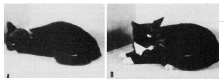
Dr. Walter Hess of the University of Zurich in Switzerland discovered that stimulation of the hypothalamus in cats can produce not only rage responses, but also fear responses. (Hess and Brügger 1943, Hess 1958)
Whereas rage responses were induced from the anterior to middle parts of the hypothalamus, fear responses were induced from the posterior part of the hypothalamus.
The stimulated cat would become restless, and move around on the experimental table, often seek a way out, and jump off the table if permitted.
First, undirected movements would be observed, which would change to escape behavior with more prolonged or intensified stimulation.
From another study. Violent escape behavior induced by electrical stimulation of the posterior hypothalamus. (Miller 1961)
A Yale University study also showed that the fear responses can occur from the posterior part of the hypothalamus in cats, confirming Dr. Hess's findings. (Roberts 1958)
The fear response induced by electrical stimulation was gradual.
At low intensities, the cat alerted, raising its head and looking rapidly around in all directions with quick, darting glances.
At higher intensities, the cat stood up and rapidly searched the entire cage, thrusting its head into corners and any opened aperture, as if attempting to escape.
After the cessation of stimulation, the activity immediately stopped.
From another study. Planned escape patterns induced from the hypothalamus. (Hunsperger and Bucher 1967)
Anterior Hypothalamus
A researcher at the University of Washington found that fear responses in cats can also occur by electrical stimulation of the anterior hypothalamus. (Nakao 1958)
On the other hand, it was the same in that the rage responses were induced by electrical stimulation of the middle hypothalamus.
Weak stimulation caused pupil dilation, crouching, looking around and a general attitude of attention or alarm. The cat sometimes mewed as if frightened or bewildered after cessation of the stimulation.
More striking responses were evocable with somewhat stronger stimulation. The pupils dilated and the eyes opened wide soon after the onset of stimulation; then, snarling, hissing, slight retraction of the ears and looking around appeared in rapid succession.
The cat finally attempted to escape from the cage. Attempts to escape were not random, but were well-directed, i.e., it consistently attacked a weak point in the cage, such as a crack or a breach in the mesh, which it attempted to enlarge by frantic clawing.
A particularly common response was leaping to the top of the cage, and while clinging to the mesh, attempting to dislodge the removable top.
Characteristically, the cats withdrew from rather than attacked the experimenter. A stick thrust into the cage was avoided even when the animal was snarling and hissing;
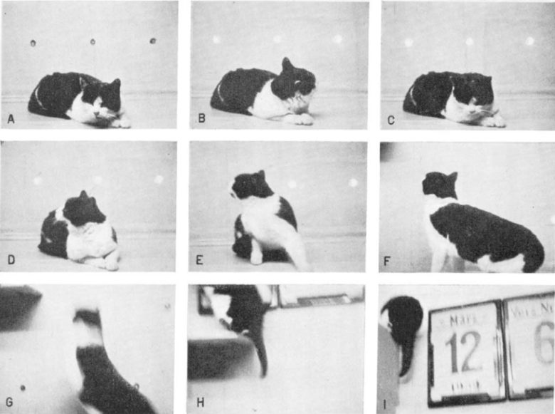
Japanese researchers also showed that fear responses in cats can be obtained from the anterior hypothalamus, confirming the previous study. (Yasukochi 1960)
It was also the same in that the rage responses can be obtained from the middle hypothalamus.
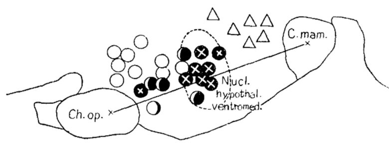
Black circle = unpleasant irritableness or rage; black circle with a cross = rage with aggression; white circle = anxiety or fear; triangle = yearning or longing.
The cats expressed typical anxiety- or fear-like responses, looking around in an apparently anxious and fearful manner with dilated pupils.
They moved around restlessly in the cage at low to middle intensities of stimulation, and with increasing intensity of stimulation, the expression of emotional agitation became more violent and hasty, and seeking around for an exit, they scratched into the corner of the cage or clawed against the mesh of it.
Menaced or agitated by the experimenter, they never tried to counterattack actively, but occasionally hissed at the experimenter, and only exhibited an aggressive attitude with a defensive posture ready to flight.
When they found a small hole in the wall of the cage, they often thrust their noses or forepaws as if they were trying to escape therethrough.
Middle Hypothalamus
The middle part of the hypothalamus has been shown to induce not only rage and aggressive responses, but also fear and flight responses.
The topographical separation is shown to be in the up-and-down direction.
Polish researchers conducted an experiment to investigate the responses induced by electrical stimulation across the anterior, middle, and posterior parts of the hypothalamus in cats. Romaniuk 1965)
They found that whereas rage and aggressive responses occurred from the ventral medial parts across the entire target regions, fear and flight responses occurred from the dorsal medial parts.
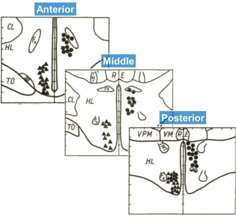
Circle = fear and flight; triangle = rage and aggression.
The stimulated cat showed pupil dilation, an increase in the respiratory rate, shrill mewing, urination, sometimes also defecation, violent motor reaction, jumping on the walls of the cage, and attempts to get out of it.
The escape attempt consisted of violently wedging the body in the cage door aperture or in the upper cover aperture and was continued all the time of the stimulation.
If only the cage door was deliberately left open, the cat instantly took its chance and jumped out of the cage, escaping in panic.
When less intensive stimulation was applied, the cat retreated to the back wall, crouched on the floor and, in such a position, remained motionless until the end of the stimulation.
When maximal stimulation was applied, the reaction of flight was chaotic, the animal rushed to and from all over the cage, running its body against the walls.

PAG
In the periaqueductal gray (PAG) of the midbrain as well, rage and fear have been observed to occur from regions that are topographically close together.
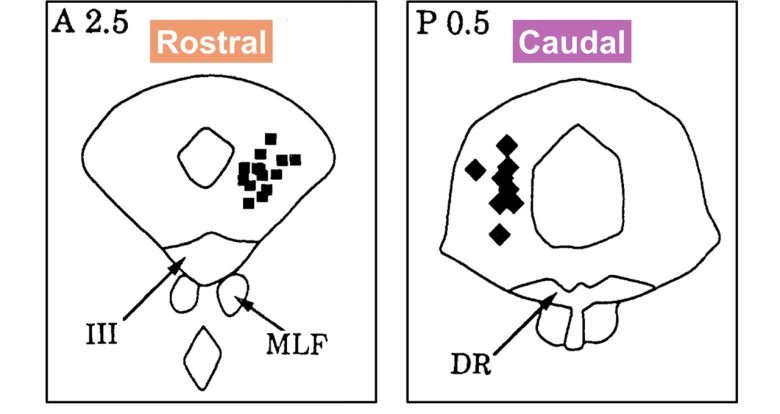
A study at the University of Nebraska showed that stimulation of the PAG in cats induced both rage and fear responses. (SKULTETY 1963)
Whereas rage responses occurred by stimulation of the rostral part of the PAG, fear responses occurred by that of the caudal part.
In the fear responses, one cat urinated, had moderate pupil dilatation, and looked quickly from side to side. It ignored being tapped in the face with a glove and attempted to escape through the open door.
Three other cats had the pupils dilated, fur standing up, the back arched, and showed urination and a clawing movement of the forelimbs.
Two hissed and growled; the other made no sound. Two ignored the glove when tapped, but the other finally bit at it after being tapped a number of times.
All cats attempted to escape when the door was opened, but they did not move around the cage, searching for a means of escape prior to this.
A University of Sydney study confirmed similar findings. (Zhang et al. 1990, Bandler and Depaulis 1991)
As in the previous study, whereas rage responses occurred by stimulation of the rostral part of the PAG, the fear response occurred by that of the caudal part.
Also, in both responses, the stimulation sites were located in the lateral and dorsolateral parts of the PAG.
In this experiment, instead of electrical stimulation, chemical stimulation was performed by injecting a glutamate analog, a neurotransmitter, into the PAG.
The cats were stimulated while restrained.
Within 5-10 seconds of the start of the stimulation, pupils dilated and fur stood up on the back and at the same time the cat usually began to struggle to free itself from restraint.
After being released, the cat began to run rapidly around the cage, moving predominantly in a direction opposite to the stimulation site, occasionally with jumps directed at the side or top of the cage.
Often the bursts of movements were interrupted by brief rest periods of 1–4 seconds.
During these brief rest periods the cat usually looked toward the top of the cage, moved its head from side to side and then often jumped.
These effects lasted for approximately 120 seconds. Afterwards, jumps or escape movements were very rarely observed.
When tested in a large room, the same stimulation evoked only running.
On reaching one end of the room, the cat usually stopped briefly, turned to the other side, and started running again. Jumps and escape movements were not observed.
Incidentally, in experiments using rodents, topographical separation of rage and fear responses has also been observed in the medial-lateral direction. (Gross and Canteras 2012, Li et al. 2025)
Amygdala
In the amygdala as well, rage and fear have been observed to occur from regions that are topographically close to each other.
A French neurologist found that electrical stimulation of the amygdala in cats can produce both rage and fear responses. (Gastaut et al. 1952)
With a weak intensity it produced an attitude of "attention," the cats opening their eyes and straightening their necks and ears.
With an increasing intensity it produced an attitude of "fear," the cats lowering their heads, sniffing, bending their ears back, and seeming to seek cover.
With an even stronger intensity it produced an "anger syndrome," the cats arching their trunks, retracting their ears, having fur standing up, and snarling.
Lateral Amygdala
Duke University researchers showed that rage and fear responses can occur from different regions by electrical stimulation of the amygdala in cats. (Shealy and Peele 1957)
Whereas electrical stimulation of the medial nucleus and central nucleus of the amygdala induced rage responses, that of the lateral nucleus and basal nucleus induced fear responses.
In the fear responses, first, the cat immediately drew back and crouched down. Then dilation of both pupils was seen. Frequently, respiration ceased for as long as 30 seconds; then escape would occur and the cat hyperventilated.
Occasionally the cat retched, sneezed, urinated, or defecated.
Throughout the stimulation, there was an attentive facial expression. Most cats were observed with their fur standing up, and whenever observed, the knee-jerk reflex was diminished during stimulation.
Swedish researchers also showed that electrical stimulation of the lateral nucleus of the amygdala in cats can induce fear responses. (Ursin and Kaada 1960)
On the other hand, electrical stimulation of the region more posterior and more medial to the fear-causing region induced rage responses.
In the fear responses, on weak stimulation, the cat immediately stopped spontaneous activities in progress, such as licking and walking ("attention"). It seemed bewildered and surprised, and behaved as if it expected something to happen.
The cat then raised the head and the upper part of the body, it looked in an inquisitive manner with quick and anxious searching movements of the eyes and head.
On increasing the stimulation intensity, the searching movements became more rapid and the cat seemed frightened.
The pupils were widely dilated and the cat cringed and withdrew a little on the platform as if prepared for flight from some unknown threat ("cowering").
The cat seemed restless and finally it jumped off the platform, ran away and hid ("flight"). In this flight the wires from the stimulator to the electrodes were automatically disconnected.
Each cat usually chose its own particular hiding place which it searched out on each stimulation.
This sequence of the behavior change, "attention" followed by "cowering" and "flight" was a typical feature of the fear responses.
Medial Amygdala
Dr. Robert Hunsperger of the University of Zurich in Switzerland showed that stimulating the medial region of the amygdala in cats can induce rage responses. (de Molina and Hunsperger 1959)
In the same region that induced the rage responses, several points were found to induce the flight responses in cats.
The primary flight responses developed in the following manner.
The pupils, after onset of stimulation, dilated, the eyes darted to and from, the head turned, occasionally fur stood up, and suddenly the cat rushed off the table.
After cessation of stimulation the excitement subsided gradually and the cat settled down, apparently relaxed.
In a few cases, however, the cat remained for a few seconds or even minutes in the posture assumed when stimulation ceased.
It looked puzzled, with pupils remaining dilated, and was indifferent to slight visual stimuli or hand clapping.
Plaintive or protesting mewing sometimes terminated this state. Flight was rarely observed after switching off stimulation.
The acoustic startle response is a biological response to sudden loud noises, and is known to be increased by fear. (Davis 1984)
A Yale University study showed that electrical stimulation of the medial nucleus of the amygdala enhanced the acoustic startle response in rats, likely as a result of the electrical stimulation producing fear in the rats. (Rosen and Davis 1988)
In the experiment, electrical stimulation of the amygdala was applied, followed by white noise as acoustic stimuli 5 milliseconds later, and the amplitude of the rats' startle responses was measured using stabilometers.
They found that electrical stimulation of several regions of the amygdala increased the acoustic startle responses by 54%-325%.
With the lowest current, the effects were observed in the region immediately medial to the amygdala, followed by the medial nucleus of the amygdala.
Hippocampus
Swedish researchers showed that electrical stimulation of the hippocampus and fimbria/fornix in cats can produce fear responses. (Kaada et al. 1953)
The fimbria and fornix are a bundle of nerve fibers that extend from the hippocampus to the septal area.
Immediately at the onset of stimulation all spontaneous activities in progress such as walking or licking ceased and the facial expression changed to "attention" or alertness.
The cat usually raised its head and upper part of the body slightly, the eyes opened, and the pupils dilated slowly, the ears pricked slightly; and there were quick searching movements of the eyes and head.
On increasing the stimulation intensity, several cats now showed signs of fear, anger, or fury during the stimulation, such as growling with gnashing its teeth, clawing, and arching their backs.
The reaction was defensive rather than aggressive in character. The cat might retire into a corner in fright, and only exceptionally did it attack the hand of the observer when approached.
Other Animals
In mammals other than cats, rage and fear have also been shown to be induced from regions that are topographically close together.
Monkeys
Hypothalamus
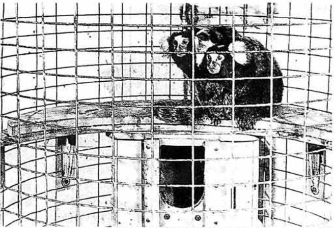
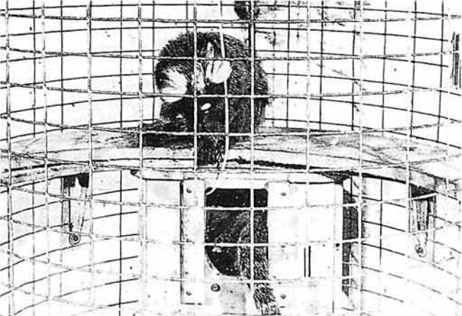
Electrical stimulation of the hypothalamus in monkeys has been shown to induce both aggressive and flight responses. (Lipp and Hunsperger 1978)
The experiment used the marmosets, and whereas electrical stimulation of the middle hypothalamus, particularly the ventromedial nucleus, induced aggressive responses, that of the posterior hypothalamus induced flight responses.
Flight responses occurred in three patterns which were interchangeably performed by the monkeys, depending on the situational context:
- "vocal threat": bursts of "ke-ke-ke…" calls
- "taking cover": entering the nest-box
- "lurking": remaining inside the nest-box
PAG
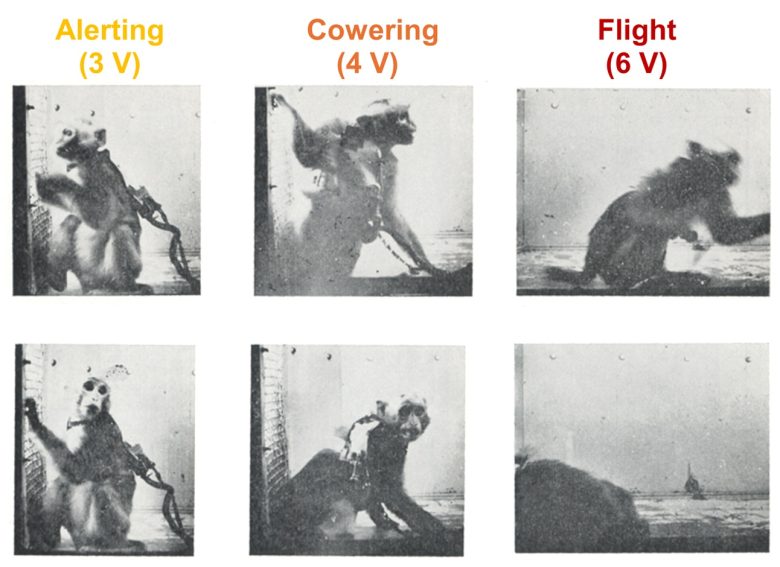
Electrical stimulation of the periaqueductal gray (PAG) in monkeys has also been shown to produce fear responses. (Segundo et al. 1955)
In the experiment, using rhesus monkeys, three types of fear responses—alerting, cowering, and flight—were induced in stages depending on the stimulation intensities.
In the alerting responses, the monkey would stop whatever spontaneous activity was in progress, raise its head, retract its ears, and become alert as if in preparation to react promptly to some unknown threat.
In the cowering responses, the monkey would cringe as if in preparation for defense or flight.
In the flight responses, the monkey would race agitatedly around the stage in what appeared to be an effort to escape an unknown but impelling danger.
Dogs
Hypothalamus
Electrical stimulation of the hypothalamus in dogs has also been shown to induce two types of responses: rage and fear. (Fonberg 1967)
In the experiment, the two types of responses occurred in the same region and could not be distinguished topographically.
In the fear and flight responses, the dog screamed, whined, stood with legs bent, tail under, or moved in all directions, tried to escape in panic, often defecated and urinated.
Heart rate was accelerated, respiration became fast and superficial.
Stimulation of the same points on different days resulted in the same behavior, which remained consistent and could be observed over weeks or months.
Rabbits
Hypothalamus
Electrical stimulation of the hypothalamus in rabbits has been shown to induce two types of responses: vigilance responses and defense responses. (Duan et al. 1996)
Vigilance responses were obtained from the dorsomedial hypothalamus, and defense responses were obtained from the lateral hypothalamus.
The vigilance responses were characterized by transient immobility, head trembling, and crouching.
The defensive responses were characterized by agitated running and hindlimb thumping, with the amount of running proportional to the stimulation intensity.
Pupil dilation and eye bulging were observed in both responses.
Amygdala
Electrical stimulation of the central nucleus of the amygdala in rabbits has been shown to induce fear responses. (APPLEGATE et al. 1983)
Stimulation yielded immediate arrest of ongoing behavior, accompanied by pupil dilation, bradycardia, and changes in breathing.
The arrest was frequently preceded by a withdrawal response from the direction of movement.
With higher intensity stimulation, vigorous running indicative of escape was immediately induced and followed by a series of hindlimb thumps.
The pattern of responses observed following stimulation was similar to that observed in response to natural threat.
Birds
Hypothalamus
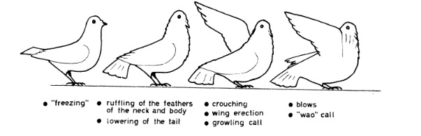
Electrical stimulation of the hypothalamus in pigeons has also been shown to induce two types of responses: defensive responses and flight responses. (Åkerman 1966)
The defensive responses were obtained from the medial region of the hypothalamus, and the flight responses were obtained from the lateral region of the hypothalamus.
In the flight responses, the pigeons tried to run or fly away swiftly almost at once at the onset of stimulation even at a low strength of current.
Rapid glances to both sides and crouching and cringing preceded the active avoidance.

In the defensive responses, the feathers became ruffled and the tail was lowered. Meanwhile the bird crouched a little, erected the wing on the opposite side from which the danger appeared and uttered a growling "alarm" call.
Powerful blows might be delivered, at first with the wing on the same side from which the danger came, and then with both wings.
In association with the blows fast cuts with the bill were delivered toward the enemy. The attacks were often accompanied by a shorter and harder vocalization sounding like—"wao."
Brainstem
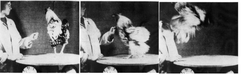
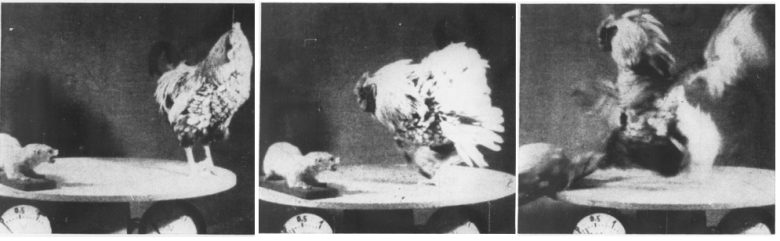
Electrical stimulation of the brainstem in chickens has also been shown to induce two types of responses: aggressive responses and flight responses. (von Holst and von Saint Paul 1962)
In the flight responses, before stimulation the rooster fed calmly.
On stimulation the rooster fixated intently on a nonexistent object approaching from a distance; the object seemed to get closer as the stimulus increased in strength and duration.
Finally the rooster jumped away fearfully. On repeated stimulation the frightening object always seemed to come from the same direction.
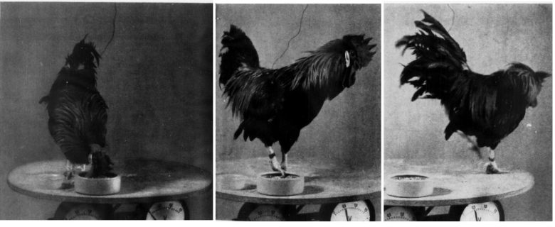
In the aggressive responses, the rooster ignored the stuffed predator before the stimulation.
When the stimulation was applied, the bird turned on the stuffed animal and attacked it furiously, spurs flying.
An attack on a friend, the rooster's keeper, was provoked if the natural enemy was absent and if the stimulation was prolonged.
The rooster preferred to aim its attack at the keeper's face rather than at her hand.