Rage
Electrical stimulation of specific sites of the brain can cause rage and aggressive behavior in humans and animals.
The three well-known sites are the amygdala, hypothalamus, and periaqueductal gray (PAG).
If abused, this would make it possible to make individuals with no violent tendency run amok and commit violent crimes.
At least in fact, experiments were already conducted more than 50 years ago to induce violent behavior in the subjects remotely and wirelessly.
Table of ContentsAll_Pages
Circuitry
Rage
The rage circuit resides in the limbic system and midbrain, beneath the neocortex, and is believed to be homologous in humans and other mammals. (Panksepp 1998)
It runs from the amygdala through the hypothalamus to the periaqueductal gray (PAG) of the midbrain.
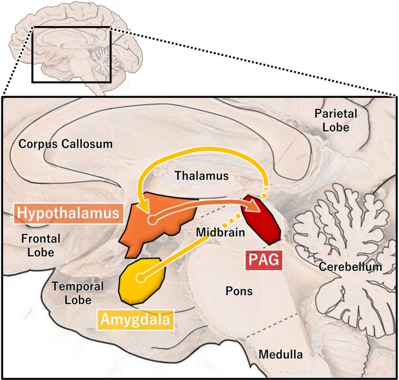

Three major brain sites related to rage. The amygdala, hypothalamus, and PAG.
(Modified from Panksepp 1998, the brain image from Vanderah 2018)
Electrical stimulation of these three regions has been confirmed to produce rage responses in both humans and animals.
If we consider the cerebral cortex as the decision-making center and the hypothalamus and midbrain as the executive centers, then the amygdala is a structure located in between. (Haller 2018)
It is generally believed that rage occurs when the amygdala is released from inhibition by the cerebral cortex and activates executive regions in the hypothalamus and PAG of the midbrain.
These regions are hierarchically arranged so that the structures at higher levels depend on the integrity of those at lower levels. (Panksepp 1998)
That is, if the PAG is damaged, electrical stimulation of the amygdala or hypothalamus will no longer produce rage responses. (de Molina and Hunsperger 1962)
However, even if the amygdala or hypothalamus is damaged, electrical stimulation of the PAG will still produce rage responses.
Aggression
Aggression is a broader phenomenon than anger itself, and aggression is not always accompanied by anger. (Panksepp 1998)
The types of aggression can be divided into two main categories: defensive rage and predatory attack, and it is only the former response that causes animals to exhibit rage responses.
There is a clear separation of neural circuits between these two aggressions.
Defensive rage, also known as rivalry aggression, involves the medial nucleus of the amygdala, the ventromedial nucleus of the hypothalamus (*), and the dorsal part of the PAG. (Haller 2013, Haller 2018)
More accurately, the mediobasal part of the hypothalamus.
Predatory attack involves the central nucleus of the amygdala, the lateral hypothalamus, and the ventral part of the PAG.
Also, the ventral tegmental area is involved in predatory attack. (Siegel and Victoroff 2009)
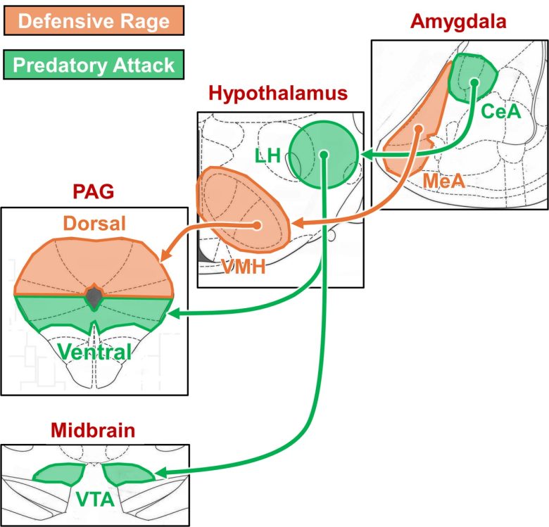
Neural circuits of defensive rage and predatory aggression
(Modified from Haller 2018, the brain atlas from Paxinos and Watson 2009)
MeA = medial nucleus of the amygdala; CeA = central nucleus of the amygdala; VMH = ventromedial nucleus of the hypothalamus; LH = lateral hypothalamus; VTA = ventral tegmental area; PAG = periaqueductal gray.
The emotions commonly referred to as "anger" or "rage" very likely derive much of their motivating energy and affective impact from the neural circuit of defensive rage. (Panksepp 1998)
In defensive rage, the sympathetic nervous system is activated, and the responses are highly impulsive, lacking the involvement of the cerebral cortex, which governs reason. (Siegel and Victoroff 2009)
The behavior occurs in nature in response to a threat by another animal of the same or different species.
On the other hand, in predatory attack, activation of the sympathetic nervous system is rarely observed. And since this response requires planning and strategy, it is thought that the cerebral cortex is involved.
In animals, the behavior is almost always directed against an animal of another species, usually a typical prey (such as a cat attacking a rat or mouse).
The predatory circuit is considered to be part of the desire circuit, and in fact, stimulating this circuit leads to feeding in some species and predatory pursuit in others. (Panksepp 1998)
For more information on the desire circuit, see "Pleasure and Desire" on page 4.
Human Predatory Attack
Some characteristics of human predatory attack is similar to those seen in animals. (Meloy 1988)
The sympathetic nervous system is minimally aroused, the target is not recognized as a threat, behavior is planned and purposeful, the attack target does not shift to another, and so on.
However, whereas in animals predatory attack is usually directed against different species, in humans it can be directed against their own species, and may be stimulated by a variety of objectives:
- The gratification of vengeful thought
- Relief from compulsive drives
- The gratification of sadistic desires
- Relief from psychotic symptoms such as paranoid delusions or auditory hallucinations
- The attenuation of pathological jealous or envious emotion
It is possible that the neural circuit of predatory atack is also used in serial and mass murders.
Humans
Electrical stimulation of the medial region of the amygdala has been shown to cause rage and aggressive behavior in humans.
This corresponds to the region that induces “defensive rage” in animals.
There is a case where stimulation of the ventral part of the PAG in the midbrain produced intense rage and aggressive behavior, even expressing murderous intent.
This corresponds to the region that induces "predatory attack" in animals.
Heath's Human Experiments
Psychiatrist Robert Heath founded the Department of Psychiatry and Neurology at Tulane University in 1949, where he conducted human experiments of electrical brain stimulation on patients with schizophrenia. (Heath 1996)
Heath states that electrical stimulation of the midbrain tegmentum, hypothalamus, medial amygdala, and hippocampus caused responses of varying intensity of fear, anger, and sometimes violence.
PAG
A male patient, "A-10," received electrical stimulation to the midbrain tegmentum, most likely the ventral part of the PAG (*). (Heath 1954, Heath et al. 1954)
This is based on the description on page 44 that "electrodes have been placed in the tegmentum of the mesencephalon a few millimeters ventral to the aqueduct," the description on page 560 that "thus far, only Patient A-10 has had stimulation to tegmental region," and the description below that the stimulation activated the oculomotor nerve.
This induced rage in the patient, and he lunged at the doctor who was present and even expressed his intent to kill him.
There remained a video that recorded this scene at Tulane University. (Berns 2005)
As the stimulation begins, the patient’s eyes flutter up and down, back and forth. (*)
This is because the stimulation activated the oculomotor nerve. (Heath 1992)
Shaking his head a few times, like a dog stung by a bee, he appeared to go into an epileptic seizure, except for the fact that he was fully conscious.
He complained of double vision and severe discomfort throughout his body, and made a claw with his right hand and began to curl up into a fetal position.
After three minutes of stimulation, he became increasingly frantic and exclaimed, “I can’t think of nothing when my brain is turning up like that. Oh, no … before I pass out! I don’t want to pass out… Oh, my brain!”
Suddenly the patient’s voice changed, and screamed in a pitch so high it was uninterpretable.
Then he started tearing at his clothes, trying to rip off his shirt, and got up from the gurney.
When the interviewer said, “You’re tearing at your clothes. Do you know you’re tearing at your clothes?” On the verge of incoherence, in a falsetto voice, the patient screamed, “I don’t care! I gotta do something! I don’t care. I don’t care!”
Pausing for a moment, he started to get off the gurney again before yelling, “I’m gonna rip you up!”
Several hands came into view and held the patient down, tying his hands.
“Stop!” the interviewer commanded. “Stop!”
The patient stared into the camera and hissed, “I don’t give a goddamn. I’m gonna kill you. Let me up. I’m gonna kill you and rip you to goddamn shreds!”
When the stimulus was turned off, the patient was perplexed by his reaction, “I have nothing against him, he was just the closest to me.” (Heath 1996)
Medial Amygdala
A 27-year-old female patient, "A-8," received electrical stimulation to the amygdala. (HEATH et al. 1955, Heath 1996)
The electrode was placed in the lateral amygdala, and this was located close to the medial amygdala.
A low-intensity stimulation to the lateral amygdala elicited a pleasurable response.
In sharp contrast, an aversive response, characterized by rage and lashing out, was observed if the current was increased sufficiently to spread and activate the medial nucleus.
As soon as the stimulating current was reduced, however, and the medial nucleus was no longer activated, the patient responded with obvious pleasure, often laughing and then, wondering aloud, “Why did I do that?”
The following is how the interview went when electrical stimulation was applied. (King 1961)
The subject's voice tone was extremely flat and lacking in emphasis. Her facial expression was blank and unchanging.
She complained of depersonalization and loss of reality, saying, "I just feel everything is all wrong. Like I can't be a part of anything. Like I didn't belong and everything is a dream or something."
A 5 mA electrical stimulation was then applied to the amygdala, and when the interviewer asked her how she was feeling, her voice became much higher in tone.
She cried, "I feel like I want to get up from this chair! Please don't let me do it!" and her facial expression altered remarkably to express pleading.
"Don't do this to me. I don't want to be mean!"
When the interviewer asked if she wanted to hit him, she replied, "Yeah, I just want to hit something. I want to get something and just tear it up. Take it so I won't!" and she handed her scarf to the interviewer.
He instead handed her a stack of paper, and without further verbal exchange she tore it to shreds.
"I don't like to feel like this!"
The level of stimulating current was then reduced to 4 mA. And she immediately changed to a wide smile, and said that she felt a little better.
When I asked her about what happened earlier, she answered, "I wanted to get up from this chair and run. I wanted to hit something; tear up something-anything. Not you, just anything. I just wanted to get up and tear. I had no control of myself."
The level of stimulating current was then increased to 5 mA again, and her voice became loud and pleading.
"Don't let me hit you!"
When asked again about how she feels, she shouted with strong voice inflection and a facial appearance of anger, "I think I feel a little better like this. I get it out of my system. I don't have those other thoughts when I'm like this…. Take my blood pressure. Make them cut this thing off, it's killing me! Take my blood pressure, I say!
"Quit holding me! I'm getting up! You'd better get somebody else if you want to hold me! I'm going to hit you!"
She raised her arm as if to strike.
The stimulating current was then reduced to 4 mA. The patient smiled widely again and laughed.
"Why does it make me do this? I couldn't help it. I didn't have any control. I wanted to slap your face. I don't like to be done like that."
Ervin and Mark's Human Experiments
Medial Amygdala
Harvard psychiatrist Frank Ervin and neurosurgeon Vernon Mark were studying many patients with violent tendencies at Massachusetts General Hospital. (Scheflin and Opton 1978, Breggin 1994)
They had been provided with the stimoceiver, a remote brain stimulation device that uses radio waves, by Jose Delgado of Yale University, and were conducting human experiments in which they induced violent behavior in the subjects remotely and wirelessly.
One of their famous subjects was Mr. Thomas R., a 34-year-old male patient, and he was a brilliant engineer with several important patents to his credit. (Mark and Ervin 1970)
Despite his muscular physique it was difficult to believe he was capable of an act of violence when he was not enraged, for his manner was quiet and reserved, and he was both courteous and sympathetic.
Mr. Thomas's chief problem was his violent rage; this was sometimes directed at his co-workers and friends, but it was mostly expressed toward his wife and children.
The assaults against his wife characteristically began after a complaint of severe abdominal or facial pain.
Electrical stimulation of the medial region of the amygdala in the subject produced the complaint of pain, and a feeling of "I am losing control."
Since they knew that both these reactions always marked the onset of one of the patient's periods of violence, the stimulation was kept short to preclude any possibility of injury.
A brief stimulation of the same site at a later date reproduced this precursory phenomenon.
Electrical stimulation of the lateral region of the amygdala, 4 millimeters sideways to the region, produced an opposite response.
Within 60-90 seconds after the onset of stimulation, it changed his mood from brooding preoccupation to relaxation.
This relaxed state did not disappear when the current was turned off, but instead dissipated slowly over a 4- to 18-hour period.
Another famous subject studied by Ervin and Mark was Ms. Julia P., a 22-year-old female patient. (Mark and Ervin 1970, DELGADO et al. 1968)
Ms. Julia had had temporal lobe epilepsy since she was 10 years old, and had frequent rage outbursts, 12 in total, some of which led to violent incidents.
On one occasion, she penetrated the heart of a girl after the girl bumped against Ms. Julia's left arm and hand in a movie theater lounge, and on another occasion, she stabbed a nurse in the lung with a pair of scissors who rushed to her aid when she showed a sign of a seizure in a hospital.
The stimoceiver was placed on her right temporal lobe, applying remote wireless stimulation to her medial amygdala.
She made a series of angry grimaces which included lip retraction and baring of the teeth, the ancient "primate threat display."
She then sprang toward the wall and attacked it.
This was sudden and unexpected, and it gave the understanding as to why victims of her attacks had not had the time to defend themselves.
The second stimulation began while Ms. Julia was playing a guitar. After 5 seconds of stimulation, Ms. Julia stopped singing and stared blankly ahead. She slipped out of communication and was unable to answer the questions posed by the psychiatrist who was examining her.
Suddenly, she swung the guitar with powerful force that it narrowly missed the psychiatrist' head and smashed against the wall.
She paced around the room for a few minutes, then gradually quieted down and resumed her usual cheerful behavior.
Both Mr. Thomas R. and Ms. Julia P. later underwent destruction of the medial region of the amygdala, which quelled their violent behavior, so Ervin and Mark reported.
However, this contradicts the claims of Harvard-educated psychiatrist Dr. Peter Breggin, who initiated his own investigation into the matter after receiving letters from a family member and a ward nurse.
Dr. Breggin found that even after the amygdala destruction, Ms. Julia's impulsive behavior persisted, and she began to deteriorate and develop suicidal thoughts, and still remained hospitalized.
As for Mr. Thomas, it turned out he was not a violent person in the first place, but rather he was unwillingly subjected to participating in the experiment on electrical brain stimulation, and then his personality was destroyed by the unnecessary brain surgery.
His violent behavior only came to happen after Ervin and Mark started intervening.
For more information, see "Early Attempts at Mind Control" on page 10.
A mild-mannered 63-year-old machinist with terminal squamous cell cancer and no particular violent history also received electrical stimulation of the medial amygdala. (Stevens 1969)
Electrical stimulation was conducted before destruction of the amygdala for the purpose of pain relief.
After the stimulation to the medial amygdala, he became wild-eyed, would not permit his nurse to come near, breathed hard, appeared extremely angry.
And He had a vacant stare, stood up in bed, attacked anyone who came near, and was incontinent of feces which he proceeded to hurl with considerable success at anyone who approached him.
This behavior continued for approximately ten minutes until a group of nurses and orderlies were able to restrain him.
The following day, the electrical stimulation was repeated for a shorter period, and a similar but milder seizure occurred.
In experiments using animals to induce rage or aggressive behavior, it is sometimes observed that incontinence occurs at the same time.
The amygdala destruction did not relieve the pain, and the patient died 7 weeks after electrode implantation.
Other Studies
Medial Amygdala
At the University of Edinburgh in the UK, coagulation of the medial amygdala was applied as a treatment for violent behavior. (Hitchcock and Cairns 1973)
Prior to the operation, 6 of the 18 patients also received electrical stimulation of the medial amygdala.
The most significant and the most dramatic effect of the stimulation was the eliciting of a range of aggressive responses.
Responses ranged from coherent, appropriately directed verbal responses (speaking to the surgeon, "I feel I could get up and bite you") to uncontrolled swearing and physically destructive behavior.
In four patients a similar pattern of restless behavior was obtained which included tearing at drapes and clothes, moving hands up toward the stereotactic frame and trying very forcibly to remove this frame.
Their actions could not be stopped by verbal commands.
Force was required in two cases to prevent the stereotactic frame from being removed by the patient.
Before and after these episodes, the two patients appeared fully aware of the situation and refrained from touching the frame or from interfering with the operative procedure in any way or from interfering with the drapes, etc.
Other than that, three patients expressed a desire to get away.
Some responses occurred within 3 seconds of the onset of the stimulation and some responses persisted 1 minute after the cessation of the stimulation.
In one of the two cases in which vehement swearing occurred, this was of sudden onset, 30-45 seconds after stimulation commenced, on three separate occasions. The disturbance lasted for 15-30 seconds and the patient became placid 5-30 seconds after cessation of stimulation.
On one occasion the patient was asked, 30 seconds after cessation of the stimulation, if he had felt angry. He agreed that he had been angry, but that he no longer was, and he sounded very surprised.
In one patient, as the result of an injury sustained during the removal of an implanted electrode, a severe right hemiplegia was produced which has resulted in a considerable disability in one arm.
Hypothalamus
At the Salpêtrière Hospital in Paris, electrical stimulation of the subthalamic nucleus was administered as a treatment to a 57-year-old man with Parkinson's disease. (Bejjani et al. 2002)
During the exploration for a valid electrode contact, stimulation of a contact in the posteromedial hypothalamus produced a sudden episode of aggressive behavior within a few seconds.
He became agitated and furious and started to shout. He aggressively attempted to get down from the surgical table while trying to take off the stereotactic frame, and had to be forcibly restrained.
During this episode, which lasted about 5 minutes, the patient exhibited a tense facial expression with bulging eyes and tachycardia.
He was vigilant, and there was no indication of confusion or seizure, but this could not be verified owing to the violence of the aggressive episode.
Stimulation was immediately stopped and a sedative was administered. After the patient awoke from sedation there was no recurrence of the aggressive behavior.
The patient recalled that he had been aggressive and that “something went wrong,” without being able to describe or explain his unexpected behavior.
Continuous electrical stimulation of an effective electrode contact in the subthalamic nucleus improved motor disability by 81% and reduced medication by 68%.
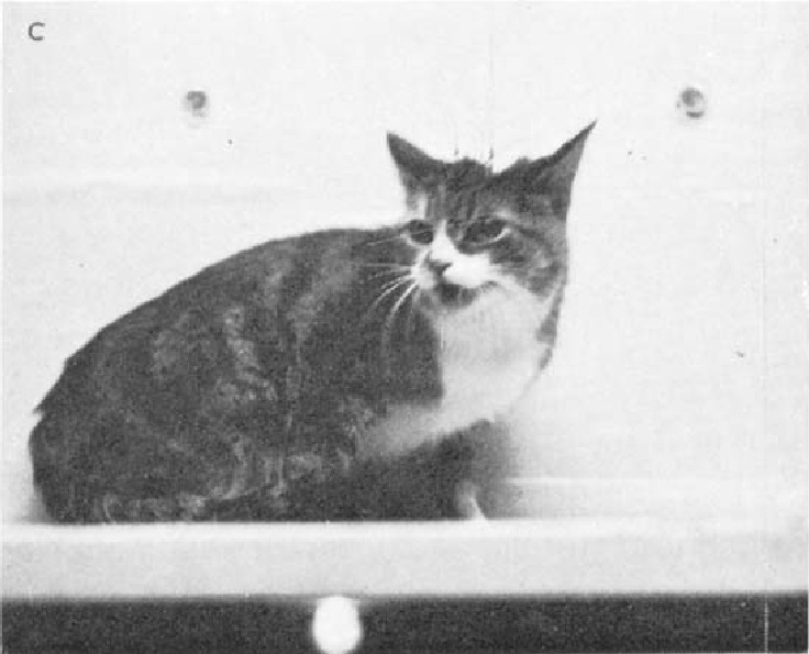
Cats
Cats are animals that have been often used to study rage and aggressive behavior.
These studies have revealed that three regions are primarily involved in rage and aggressive behavior: the amygdala, hypothalamus, and periaqueductal gray (PAG).
Hypothalamus
Dr. Walter Hess, a physiologist at the University of Zurich in Switzerland, began research into mapping the functions of the diencephalon (thalamus and hypothalamus) in the 1930s, and was awarded the Nobel Prize in Physiology or Medicine in 1949.
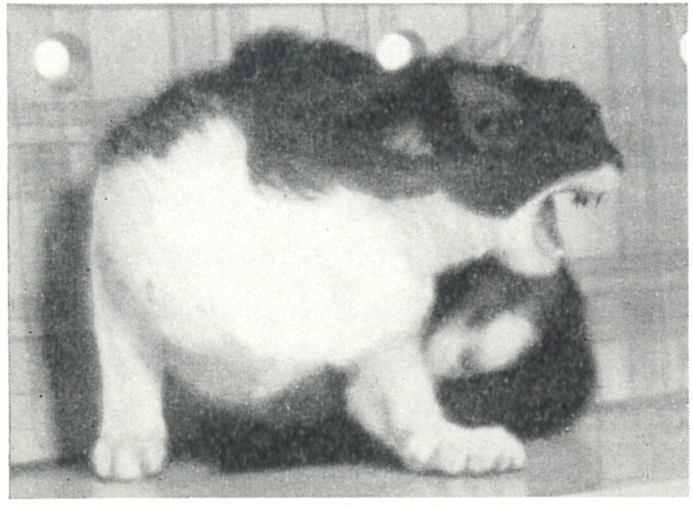
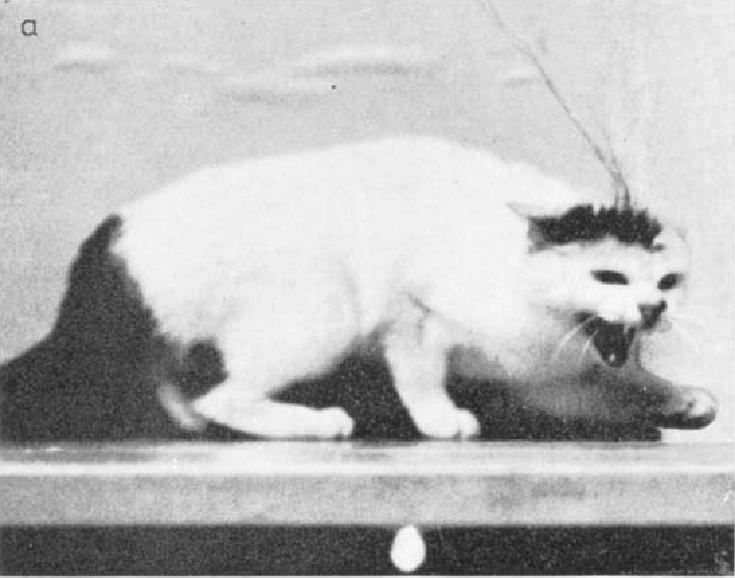
In the course of his research, he discovered that electrical stimulation of the hypothalamus in cats can produce rage and aggressive behavior. (Hess and Brügger 1943, Hess 1958)
The stimulated cat would act as if it were being threatened by a dog.
The cat would spit, snort, or growl; fur would stand up on the back; the tail would become bushy; the pupils would widen sometimes to their maximum; and the ears would lie back or move back and forth.
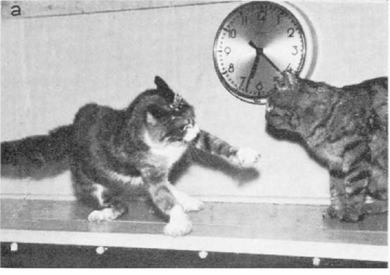
When the stimulation was maintained or intensified, the cat would make an actual attack. The cat would turn toward a person standing in its vicinity and leap on him or strike a well-aimed blow at him with its paw.
In such an excited state, it was not uncommon for the cat to expel feces, and more frequently, spurt urine.
The behavior ceased as soon as the electrical stimulation ceased, but the cats remained irritable for several minutes and became hostile when the researchers approached them.
From another study: electrical stimulation of the hypothalamus induced an emotional defensive response. (Akert 1961)
Many researchers were inspired by Dr. Hess's experiments, and conducted the experiments that induce rage and aggression by electrical brain stimulation.
Neurophysiologist Jose Delgado of Yale University is one of them. (Delgado 1964)
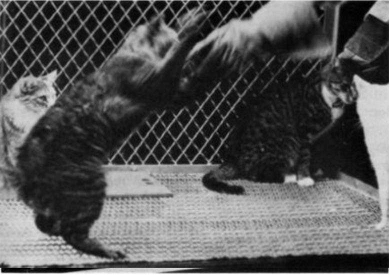
When the lateral hypothalamus was stimulated, the cat showed the aggressive attitudes with the pupils widening, fur standing up, hissing, showing of the teeth, snarling expression, and other manifestations.
The cat then advanced, directing well-aimed blows with unsheathed claws against other cats, which fought back or withdrew.
Attempts by the investigator to protect the attacked cats diverted the attack of the stimulated cat, which bit and clawed the gloved hand ferociously.
The aggressive behavior usually started and ended with the stimulation, but distrust toward the experimental cat persisted.
Even if the stimulated cat was small and was struck and clawed by the others, attacks and fights were elicited every time the hypothalamus was stimulated. A painful experience did not inhibit evoked aggressiveness.
The cats also disliked the stimulation and learned to stop the brain stimulation by turning the pedal.
The Ventromedial Nucleus of the Hypothalamus
Subsequent researchers looked more closely at the sites in the hypothalamus that produce rage and aggression responses.
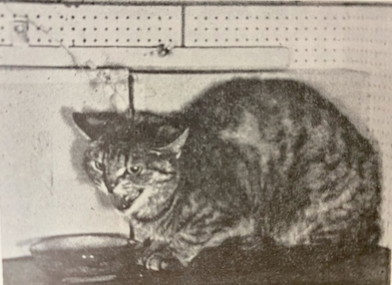
A study at the University of Washington showed that electrical stimulation of the middle hypothalamus in cats, specifically the ventromedial part, can produce aggressive behavior. (Nakao 1958)
Typical reactions included pupil dilation, retraction of the ears, arching of the back, and extension of the legs.
With weak stimulation, spontaneous behavior patterns, such as drinking milk, grooming, purring and walking, stopped abruptly.
The pupils dilated and slight snarling occurred, but if moving objects or an experimenter were not in view, the responses did not progress further.
However, when the experimenter was visible, the cat retracted its ears, arched its back, and snarled and hissed at him.
With stronger stimulation, any slight movement by the experimenter provoked a violent and directed attack. When a stick was offered, the cat bit it fiercely.
In a study conducted at Kyushu University in Japan, most aggressive behavior in cats was produced by electrical stimulation of the ventromedial nucleus, located in the middle hypothalamus. (Yasukochi 1960)
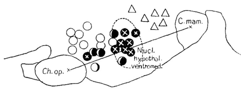
Black circle = unpleasant irritableness or rage; black circle with a cross = rage with aggression; white circle = anxiety or fear; triangle = yearning or longing.
The cats were only staring around and growling unpleasantly during less intensive stimulation.
The cats were agitated gradually and became more aggressive with increasing intensity of stimulation, with pupils widely dilated, ears retracted back, fur standing up, hissing with teeth threateningly exposed, and finally sprang and rushed against the agitating experimenter with furious growling.
In some cases cats attacked fiercely at a harmless corner of the cage, as if they had a visual hallucination of some hostile object.
PAG
Electrical stimulation of the periaqueductal gray (PAG) of the midbrain has also been shown to produce rage and aggressive responses.
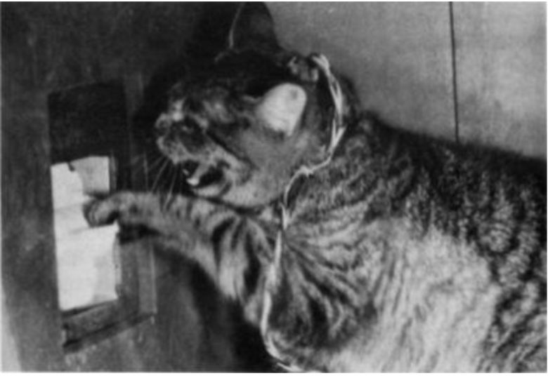
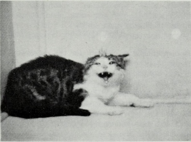
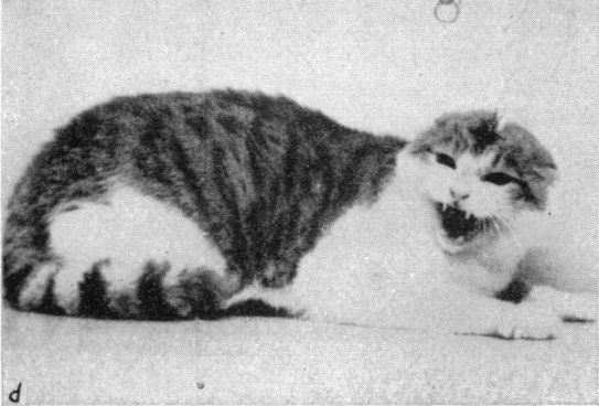
Dr. Robert Hunsperger of the University of Zurich discovered that electrical stimulation of the PAG in cats can induce rage and aggressive responses. (Hunsperger 1955)
The rage reactions upon stimulation developed as follows: first lid widening, retraction of the third eyelids and pupil dilatation appeared.
Excitement then set in: the pupils dilated more and more, sometimes maximally, and the cat gazed fixedly at some invisible foe. It hissed suddenly and repeatedly, baring its teeth. Growls sometimes accompanied the outburst.
After increased stimulation the cat passed on to attack a person or a stick put in its way.
These rage and aggressive responses occurred more rapidly from the PAG than from the hypothalamus.
A researcher at the University of Nebraska also showed that electrical stimulation of the PAG in cats can induce rage and aggressive responses, confirming Dr. Hunsperger's findings. (SKULTETY 1963)
The stimulated cats showed these general characteristic rage responses: the ears flattened, the back arched, the fur standing up on the back and tail, the pupils dilated, hissing and growling, and urinating frequently.
If a glove was introduced into the observation box, the cat would launch a well-directed attack.
Although the response stopped as soon as the stimulus was turned off, most of the cats subsequently licked their lips, mewed, and paced around restlessly for 15 to 30 seconds.
Amygdala
Electrical stimulation of the amygdala has also been shown to produce rage responses, but this does not seem to lead to aggression as much as other sites.
A French neurologist found that electrical stimulation of the amygdala in cats can produce rage responses in the cat. (Gastaut et al. 1952)
With a weak intensity, it produced an attitude of "attention," the cats opening their eyes and straightening their necks and ears.
With an increasing intensity, it produced an attitude of "fear," the cats lowering their heads, sniffing, bending their ears back, and seeming to seek cover.
With an even stronger intensity, it produced an "anger syndrome," the cats arching their trunks, retracting their ears, having their fur standing up, and snarling.
The anger was undirected and the cats were less sensitive than normal to external stimuli during the electrical stimulation.
Medial Amygdala
A Duke University study showed that within the amygdala in cats, electrical stimulation of the medial nucleus and central nucleus produces rage responses. (Shealy and Peele 1957)
The stimulated cats snarled, reared up on their hind legs, unsheathed claws, pawed the air, and tried to bite or scratch anything in the vicinity.
The rage was not directed at any particular person or object.
In addition, autonomic responses such as salivation and nasal secretion, vomiting, sneezing, urination, and defecation were occasionally induced.
Dr. Hunsperger focused not only on the amygdala but also on nerve fibers called the stria terminalis to closely investigate the sites that induce rage responses in cats. (de Molina and Hunsperger 1959)
The stria terminalis is a bundle of nerve fibers that serves as the main output pathway for the amygdala, extending from the medial part of the amygdala to the ventromedial nucleus of the hypothalamus.
As mentioned above, the ventromedial nucleus of the hypothalamus is the region that induces rage and aggressive responses.
The electrodes that induced rage responses were found to be concentrated in the medial region of the amygdala, where the stria terminalis originates, which corresponds to the region adjacent to the medial nucleus and central nucleus, and the dorsal part of the basal nucleus.
Electrical stimulation of the stria terminalis itself also induced rage responses.

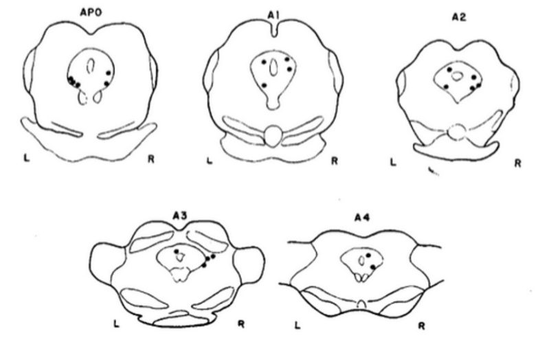
The sites of the amygdala and stria terminalis that induced rage responses.
Black circle = threat with growling; black square = threat with growling followed by hissing; white circle = flight of unpremeditated character. (Hunsperger and Bucher 1967)
The cat, before the onset of stimulation, sat or lay quietly on the table.
Soon after stimulation set in, the cat turned its head toward the observer, the lids widened, the pupils dilated and respiration increased in depth and rate. The eyes remained fixed on the observer and the cat appeared to be taut.
A first low growl was then emitted, chin protruded and upper lip pursed, followed by louder growls uttered at intervals of 5-10 seconds.
Often the pupils dilated maximally, the ears were laid back, the head was lowered, and fur stood up on the back and tail.
The growling sometimes alternated with short puffing expirations during which the cat stood up and slightly hunched its back.
When the response elicited was growling followed by hissing, toward the end of stimulation the cat became to repeat raucous hissing; the mouth was opened wide, with corners retracted, teeth bared.
Although the cat turned its head toward the observer, the cat never attacked.
Depending on the sites stimulated, the cat may end up running away.
Differences between the Sites
Dr. Hunsperger noticed that electrical stimulation of the hypothalamus, amygdala, and PAG of the midbrain in cats each induced different responses. (Hunsperger and Bucher 1967)
At the hypothalamus, the response strikingly resembled the natural reaction, seen when a cat meets a dog, or an unfriendly fellow cat.
The cat raised a forepaw, ready to strike. Vigorous hissing accompanied the threatening display. Sometimes, after a fierce threat display, the cat resorted to flight.
At the amygdala, the growling accompanied the defensive display. Hissing only occurred after prolonged stimulation.
The response, in contrast to the reaction obtained from the hypothalamus, showed less "acting out."
At the PAG of the midbrain, it always produced hissing and pupil dilatation, but less regularly a full threat pattern.
However, the time it took the cats to hiss after stimulation was shorter than at the hypothalamus.
In addition, when the hypothalamus and PAG were stimulated, the cats engaged in aggressive behavior when a stick or a human hand was brought close to them.
When a stuffed cat was placed on the table during stimulation of the hypothalamus, the defense pattern, threat followed by occasional flight, was modified.
Sometimes the excited cat, hissing, with claws unsheathed, warded off the foe. A moment later, it jumped from the table in a frenzy. At other times, the cat, still hissing, with claws unsheathed, struck the dummy’s face.

That stimulation of the PAG of the midbrain produced the incomplete patterns may be due to stimulation of points that induced fragmented responses rather than points that induce integrated aggressive behavior.
In fact, an experiment using monkeys has shown that aggressive behavior can be broken down into fragmented movements at the brainstem level, including the midbrain. (DELGADO 1967)
Defensive Rage and Predatory Aggression
Hypothalamus
Psychologist Dr. John Flynn of Yale University discovered that there are two types of aggressive behavior induced by electrical stimulation of the hypothalamus in cats. (WASMAN and FLYNN 1962)
One is defensive rage induced by the medial hypothalamus, and the other is predatory aggression induced by the lateral hypothalamus.
Defensive Rage
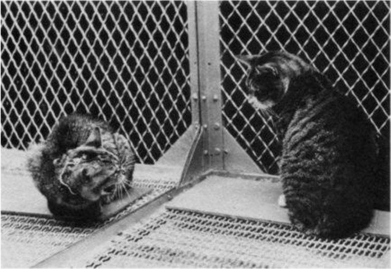
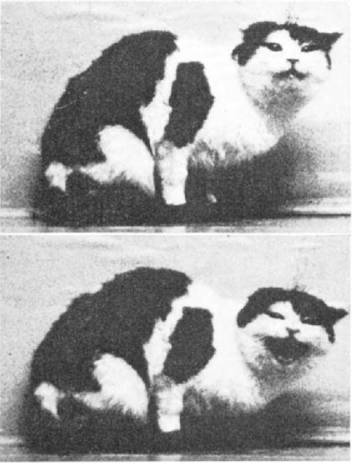
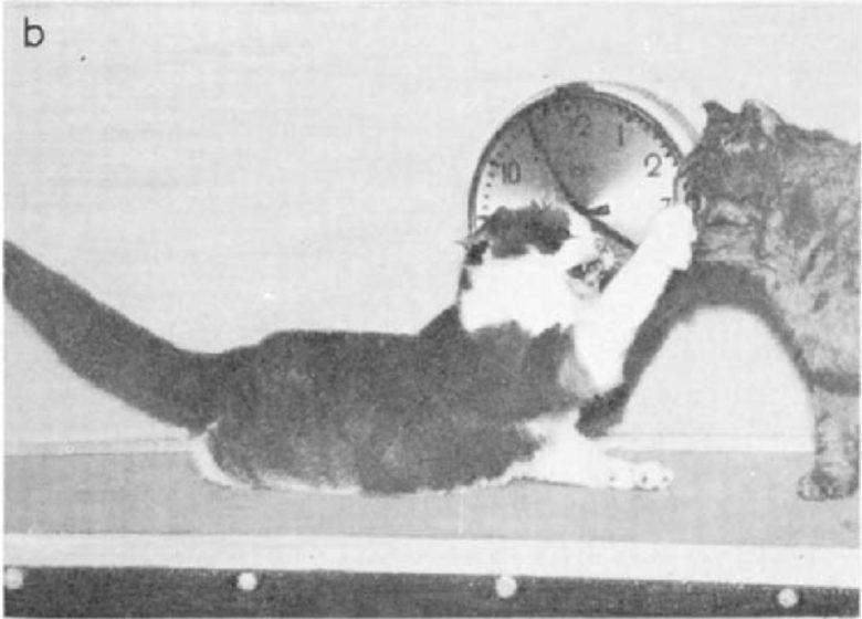
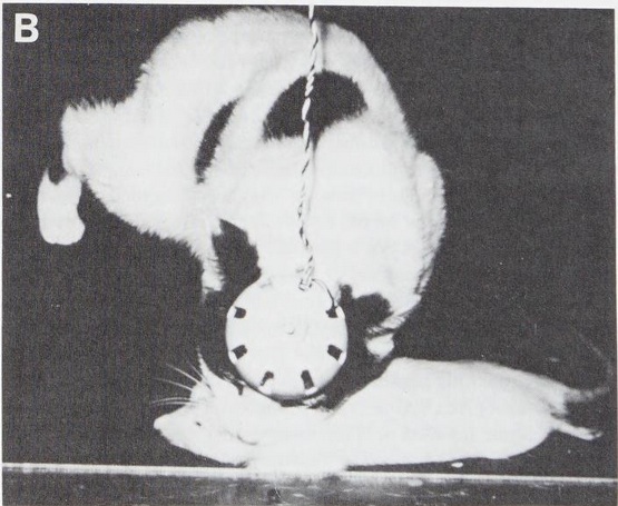
A cat raged toward a rat with electrical stimulation of the medial hypothalamus. (Flynn 1967)
This attack was characterized by a pattern of pronounced sympathetic arousal commonly regarded as indicative of cats' rage. The response invariably began with behavioral alerting and dilation of the pupils.
With weaker stimulation, the cats just remained alert even after two minutes of stimulation.
With stronger stimulation, in addition to the initial alerting, fur stood up noticeably and the tail became bushy and fluffed out.
Hissing occurred alternating with low growls. Urination often occurred on the first trial of a session. If sitting, the cat leaped to its feet and began to move with head low to the ground, back arched, claws unsheathed, hissing and snarling, salivating profusely, and breathing deeply.
The cat stood poised, appearing to watch the rat intensely, while the affective aspect of the reaction became still more pronounced.
After a second or two, the cat raised a paw with claws unsheathed and then struck with its paw in a series of swift, accurate blows. Any sudden movement of the rat served to trigger the attack, but the attack would occur even if the rat remained motionless or frozen.
Predatory Attack
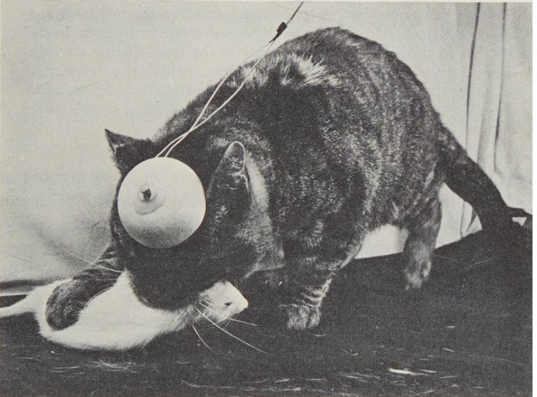
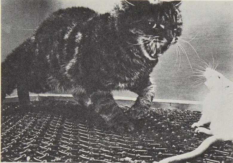
A cat stalked a rat and bit it on the neck with electrical stimulation of the lateral hypothalamus. (Flynn 1967)
The second form of attack pattern reminded observers of an animal stalking its prey.
With weaker stimulation, the cat usually leaped to its feet almost with the onset of the stimulation and attentively looked around from side to side. If the stimulation were continued, it would walk around the cage but would ignore the rat.
With stronger stimulation, the cat moved swiftly with its nose low to the ground, back arched, and hair slightly on end. Sometimes the cat's nose twitched slightly, as if it were sniffing.
The cat usually went directly to the rat and bit viciously at its head and neck.
The cat used its paws primarily to knock the rat on its back in order to get at its throat. The paws were used neither to deliver discrete blows nor to rake the rat with its claws.
Although the cat would sometimes bite the rat's stomach or back first, biting was ultimately aimed at the head and neck.
On some trials the cat circled around the observation box a few times ignoring the rat or sniffing at it in passing. In the course of circling the cat would suddenly pounce on the rat and savagely tear at it with its mouth.
The predatory attack was characterized, especially at weak stimulation, by only minimal signs presumed indicative of cats' rage.
The cats rarely hissed and never growled, emitted high-pitched screams, nor salivated profusely. The cat's movement around the cage, however, was quicker and more persistent.
Although this predatory attack was never as dramatic as defensive rage, it was, in reality, more effective and deadly, as far as the rat was concerned.
The Differences between Defensive Rage and Predatory Attack
The attack responses were completely under stimulative control in the sense that the attacks ceased precisely with the offset of the stimulation.
A swipe with the paw was carried through if sufficient momentum had been gained, but a bite could be halted just as the jaws were closing.
The experiment involved the use of docile cats that would not spontaneously attack rats, but when brain stimulation was given, they began to aggressively attack the rats.
Defensive rage was aversive to cats and they would avoid it if given the opportunity, but cats did not show an aversive reaction to predatory attack. (Adams and Flynn 1966)
Rather, predatory attack was rewarding for the cats, and the docile cats that normally would not attack rats began to prefer to enter the room where they could attack rats after receiving brain stimulation. (Roberts and Kiess 1964)
Ventral Tegmental Area
Dr. Flynn also discovered that predatory aggression in cats can also occur by electrical stimulation of the ventral tegmental area. (Bandler et al. 1972)
This predatory attack resembled that induced by stimulation of the lateral hypothalamus.
When stimulation began, the cat moved swiftly with its nose low to the ground and hair slightly on end and went directly to the rat and bit its head and neck repeatedly, usually killing it with the first few bites.
Although the cat would sometimes bite the rat's tail, stomach, or back, biting was ultimately directed to the neck and head.
On some trials the cat would circle the test cage several times, ignoring the rat, and then suddenly pounce on the rat and bite it savagely.
This attack was not accompanied by signs of autonomic arousal other than that the pupils dilated and the fur stood up along the tail and midline of the back.
The lateral hypothalamus and ventral tegmental area are thought to be part of the desire circuit.
In fact, it has been confirmed that stimulation of the lateral hypothalamus and ventral tegmental area not only induces predatory attack, but also induces motivated behavior such as eating, drinking, and mating. (See page 4, "Pleasure and Desire.")
Amygdala
Dr. Flynn also found that predatory aggression was facilitated by electrical stimulation of the lateral nucleus of the amygdala. (Egger and Flynn 1963)
In the experiment, electrical stimulation of the lateral hypothalamus was first delivered to induce predatory attack.
Some attacks were characterized by biting, others by striking with the paw. Mixed forms with both biting and striking occurred.
Electrical stimulation of the lateral nucleus of the amygdala then facilitated this predatory attack.
To be specific, the cats came to launch attacks more rapidly.
A subsequent study has shown that the central nucleus as well as the lateral nucleus of the amygdala facilitates predatory attack. (Block et al. 1980)
On the other hand, defensive rage has been shown to be facilitated by electrical stimulation of the medial nucleus and basomedial nucleus of the amygdala. (Stoddard-Apter and MacDonnell 1980)
In the experiment, electrical stimulation of the ventromedial nucleus of the hypothalamus was first delivered to induce defensive rage.
The cats hissed, had fur standing up, dilated their pupils, retracted their ears back, and growled.
Electrical stimulation of either the medial or basomedial nucleus of the amygdala then facilitated this defensive rage.
To be specific, the cats came to hiss more rapidly.
PAG
The distinction between defensive rage and predatory attack was also found to exist in the PAG of the midbrain. (Bandler 1975, Bandler 1977)
When electrical stimulation was given to the dorsal part of the PAG, the cats manifested rage.
The cats hissed, spit, growled, bared the teeth, laid back the ears, arched the back, and struck repeatedly with the paws.
On the other hand, when electrical stimulation was given to the ventral part of the periaqueductal gray matter, the cats began to prey.
The cat moved swiftly with its nose low to the ground, went directly to the rat, and bit its neck and head repeatedly, usually killing it with the first few bites.
This attack was not accompanied by signs of autonomic arousal other than that the pupils dilated and the fur stood up along the back and tail.
Brain Map of Aggression
Hypothalamus
Dr. Allan Siegel of the University of New Jersey Medical School also conducted experiments to induce two types of aggressive behavior in cats, confirming the previous research. (Siegel and Brutus 1990)
Similarly, in his experiment, electrical stimulation of the medial hypothalamus in cats induced defensive rage, while that of the lateral hypothalamus induced predatory attack.
He expanded on Dr. Flynn's research, investigating the regions of the hypothalamus in cats where attacks are induced with greater precision, drawing a detailed brain map.
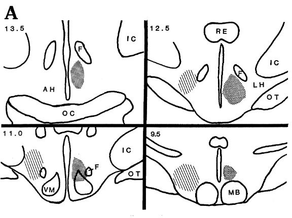
VM = ventromedial nucleus; F = fornix; MB = mamillary body; LH = lateral hypothalamus; OT = optic tract; RE = reuniens nucleus of thalamus; IC = internal capsule.

PAG
Dr. Siegel also confirmed the previous research on two types of aggression in cats regarding the PAG of the midbrain. (Shaikh et al. 1987)
Similarly, in his experiments, electrical stimulation of the dorsal part of the PAG induced defensive rage, while that of the ventral part induced predatory attack.
He also investigated the regions of the PAG in cats where attacks are induced with greater precision, drawing a detailed brain map.
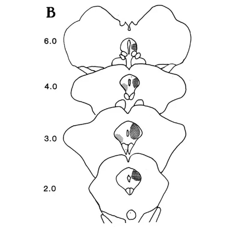
The brain map of aggression in the PAG. Stippled regions (dorsal parts) induce defensive rage whereas striped regions (ventral parts) induce predatory attack. (Siegel and Brutus 1990)
Monkeys
Several experiments have been conducted by a limited number of research groups to induce rage and aggressive behavior in monkeys using electrical stimulation.
Rather than exploring neural circuits, many of the experiments focus on changes in behavior in social situations.
These experiments of electrical brain stimulation on monkeys are said to have raised suspicions of mind control in human society. (Blackwell 2014)
Delgado's Experiments
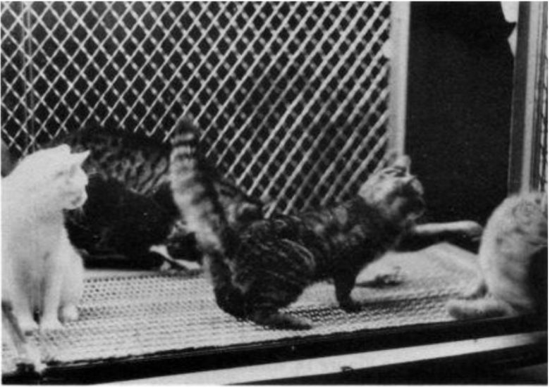
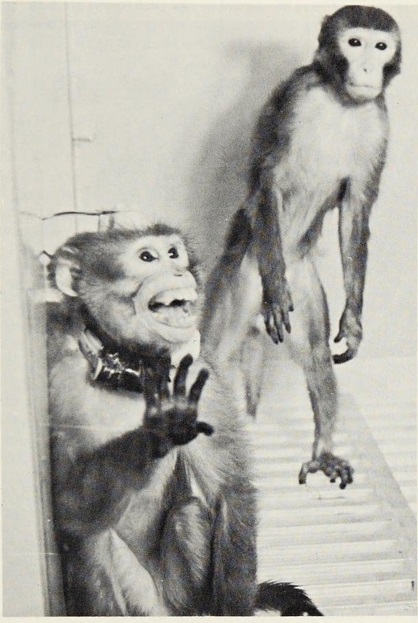
Neurophysiologist Jose Delgado at Yale University conducted the experiments of electrical brain stimulation on monkeys living in colonies to study the effects on their behavior in social situations. (Delgado 1964)
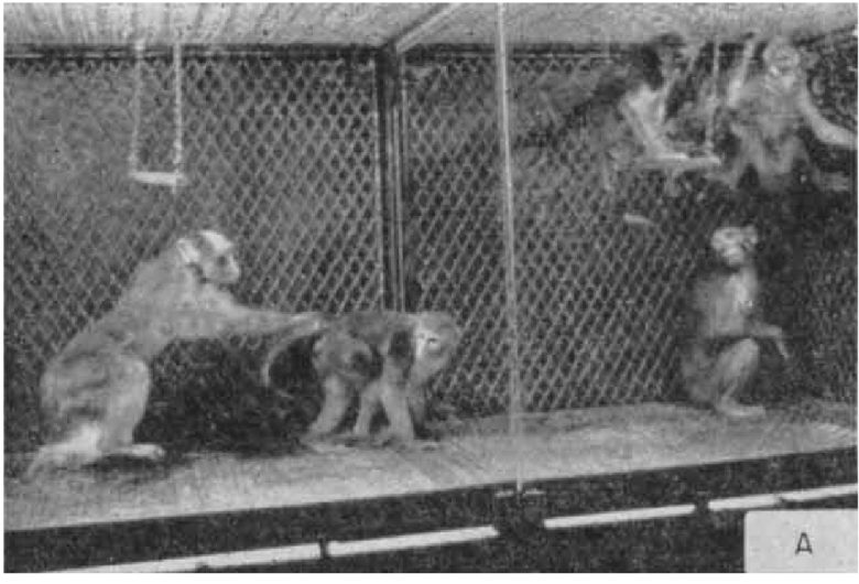
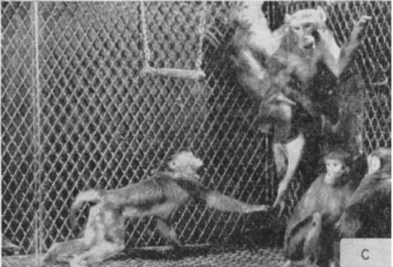
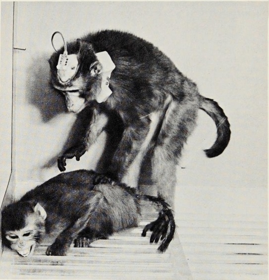
Electrical stimulation of the PAG in the boss monkey produced an immediate increase of aggressiveness.
The boss ran around the cage and attacked the other members of the group, chased them, and even bit them if they were unsuccessful in eluding him.
The pattern of the aggressive behavior was the same as spontaneous attacks, but was more violent and persistent, and lasted as long as the stimulation was applied.
The attacks were well-aimed and well-organized, and showed social selectivity, because they were directed especially toward monkey no. 4, a male with whom the boss had a previous hostile relationship.
The increase of evoked aggressiveness in the boss was also manifested sometimes by biting the swing, shaking the cage, and attacking its mirrored image in the glass front of the cage.
There was also a social spread of aggressiveness.
Each time the boss monkey received electrical stimulation, a friendly and intimate female monkey no. 7 joined him in attacking the rest of the colony.
In a few cases, the attacks were even launched by female monkey no. 7, in spite of the fact that she was much smaller than the other monkeys.
These friendly associations and the selection of the animal to be attacked were determined by previous relations within the colony, and replicated past spontaneous groupings and clashes. (Delgado 1967)
Brain stimulation did not modify social likes and dislikes, but only increased the number of aggressive behavior.
Therefore, electrical brain stimulation was considered to have changed the emotional state of the animals rather than induced stereotypical movements during attacks.
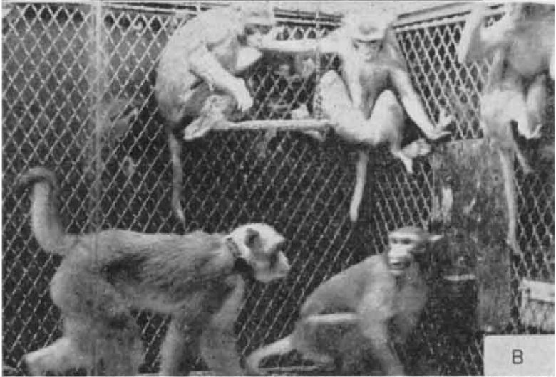
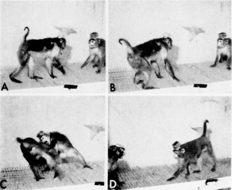
When a boss monkey received electrical stimulation to its thalamus, it threatened monkey number 3 100% of the time. The photo is from another Delgado's study. (DELGADO 1966)
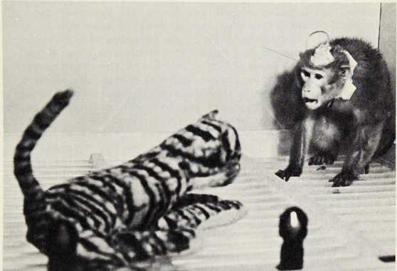
Delgado also showed that the aggressive responses elicited by electrical brain stimulation depended on the monkeys' social rank. (Plotnik et al. 1971)
However, it should be noted that the sites stimulated in this experiment were the thalamus and trigeminal nerve, so this was probably secondary aggression caused by pain.
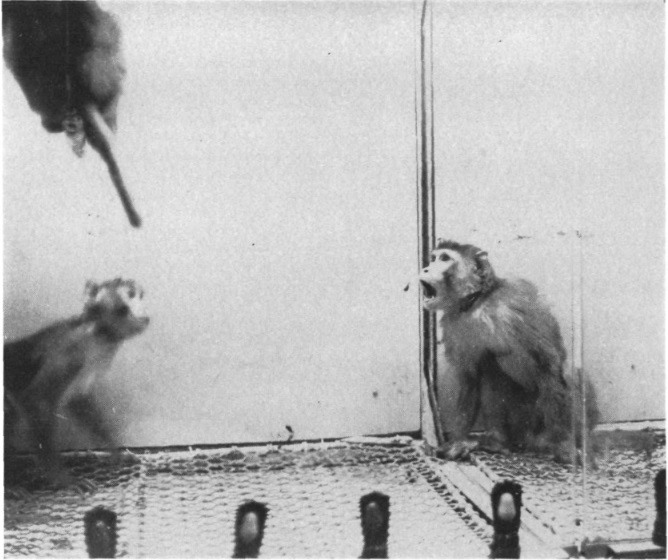
For example, monkey A6 did not threaten or attack the dominant monkey after electrical stimulation, but instead simply grimaced.
On the other hand, A6 threatened and attacked the subordinate monkey and, more frequently, a toy tiger.
Yerkes National Primate Research Center Experiments
The Yerkes National Primate Research Center also conducted the experiments of electrical brain stimulation on monkeys living in colonies to study the effects on their behavior in social situations. (Robinson et al. 1969)
In an experiment, electrical brain stimulation was shown to change the rank of a monkey colony.
The groups consisted of a dominant male, a subordinate male, and a female. The dominant monkey paced around the cage, displaced or actively assaulted the subordinate, and monopolized the female.
Electrical stimulation of the hypothalamus in the subordinate male resulted in attacks directed against the dominant monkey, who fought back vigorously.
Attacks were not made against the female or against inanimate objects.
The attacks usually occurred within 5 seconds after the onset of stimulation and fighting usually ceased after the termination of the stimulation, but occasionally the fight continued by the dominant monkey.
These fights were intense and vicious, resulting in cuts and loss of hair.
Initially the subordinate monkey lost most of the fights, and in between stimulations he continued to make submissive gestures.
With repeated stimulation of the subordinate monkey the dominant monkey began to fight less effectively and to sit quietly in between stimulations.
Following the 10th stimulation in one group and the 43rd stimulation in the other, the 2 males reversed status positions.
With the approach of the change the initially dominant monkey began to give loud, shrill screams each time he was attacked.
Following the dominance reversal, brief weak stimulations which produced only threat behavior in the stimulated monkey sufficed to produce fear vocalizations and flight in the threatened monkey.
The newly dominant male now paced freely around the cage, displaced his opponent easily, mounted the female and eventually was groomed by her.
Another experiment at the research center investigated the effects of sex and dominance on induced aggressive responses in monkeys. (Alexander and Perachio 1973)
In the experiment, they first confirmed that electrical stimulation of the hypothalamus in monkeys induced aggressive responses.
Weak electrical stimulation caused the monkeys to threaten, and increasing the intensity of the stimulation caused the threats to turn into attacks.
When the attack occurred, it was followed by fighting behavior such as biting, slapping, and holding.
Next, the effects of sex and dominance on aggression in the monkeys were examined.
In a situation where both dominant and subordinate monkeys were present, when given electrical stimulation, the experimental monkeys prioritized attacking the subordinate monkeys.
In a situation where only dominant monkeys were present, the experimental monkeys either failed to attack or attacked less frequently.
Males were also attacked preferentially over females.
In a situation where only females were present, however, it was demonstrated that stimulated males can attack females quite easily.
Other Experiments
Dr. Robert Hunsperger of the University of Zurich showed that, as in cats, aggressive responses can also be induced in monkeys by electrical stimulation of the ventromedial nucleus of the hypothalamus. (Lipp and Hunsperger 1978)
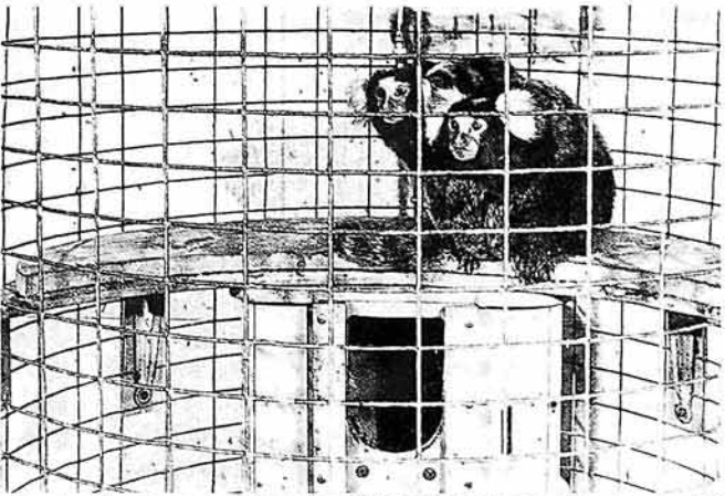
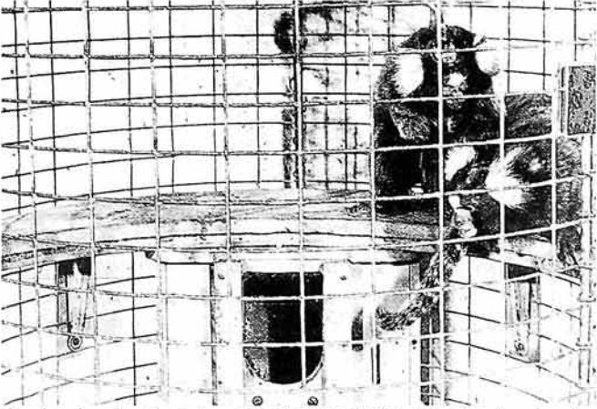
The experiment used the marmosets, and the attack response consisted of the following elements:
- "Vocal threat": bursts of "ke-ke-ke…" calls
- "Short attacks": occasional grabbing, slashing or biting of either the partner or an inanimate target
- "Violent attack": quick movement over some distance (at least 12 in [30 cm]), followed by sustained slashing, grabbing or biting, and chasing of the target
- "Fighting": mutual grabbing, slashing and biting of two monkeys

Other Animals
In other animals, electrical brain stimulation has been shown to produce two distinct types of aggressive responses as well.
Opossums
In opossums as well, electrical stimulation of different sites of the hypothalamus has been shown to produce two distinct aggressive responses against rats. (Roberts et al. 1967)
Stimulation of the medial hypothalamus induced defensive threatening behavior.
The opossum opened the mouth wide, growled, swung the head and shoulders from side to side, and backed.
He tended to back into a corner where his rear was protected and the open jaw was swung in an arc to the front, creating a formidable defensive posture.
Despite its aggressive appearance, the response was almost purely defensive.
Whether tested with a stuffed opossum, a live opossum, or a stick moved near and in the mouth, actual biting was infrequent and brief and not directed appropriately.
Stimulation of the lateral hypothalamus induced predatory attack.
The primary components of the attack pattern were seizing, head tossing, kill biting, and crunching.
The opossum fixed his gaze on the rat and began to approach with neck extended.
After capturing the rat in its mouth, the opossum violently tossed its head, causing damage to the rat.
Between the head-tossing, the opossum gave the rat several lethal bites, which were repeated as long as the rat continued to resist.
When the rat ceased to struggle, the head-tossing dropped out, and the biting was shifted to the head where it became rhythmic and caused an audible crunching.
The more energetic rats were often able to fend off the opossums for short periods, but when the intensity of the stimulation was increased, the attack tendency of the opossums became stronger, and the rats' resistance ended up futile.
The attack always ended immediately after the stimulation ended.
The stimulation even produced predatory attack against 60-70 day old opossum pups, which were brutally eaten like mice.
Spontaneous predatory attack was similar to those induced by stimulation, but had a more gradual onset, were less intense, and were sometimes mixed with temporary retreat.
Rats
In rats alike, electrical stimulation of different sites of the hypothalamus has been shown to produce two distinct aggressive responses.
Electrical stimulation of the ventromedial nucleus of the hypothalamus and its dorsolateral region produced defensive rage. (Kruk et al. 1983)
Aggression consisted mainly of attack jumps and bite attacks.
In attack jumps the stimulated male rat approaches another male and jumps upwards in front of the latter.
While jumping it tries to bite the head of the opponent and at the same time it tries to deliver a fierce jump in the belly of the usually upright defensive opponent.
In bite attacks the stimulated rat simply bit the opponent in the head without jumping.
Either attack may be followed by a fierce clinch-fight.
Electrical stimulation of the lateral hypothalamus produced predatory atack against mice and juvenile rats. (Woodworth 1971)
The primary components of the attack were seizing, attack jumping, and kill biting.
The seizing bite, accompanied by grasping with the forepaws, was more often directed to the anterior part of the body.
Attack jumping occurred after the seizing bite, and took place so rapidly that it could only be followed in the high-speed films.
The target continued to be held with the mouth and paws during the jumping, and the rat often kicked the target with its hind legs while in the air.

Kill biting consisted of repeated bites that were shifted all over the body.
The head was thrust forward with each closing of the jaws, and the forepaws were used to pin the attack object to the floor or to change its position in the mouth.
Biting always ceased within a second of stimulative offset, although the targets were not always immediately dropped.
Dogs
In dogs as well, electrical stimulation of the hypothalamus has been shown to produce rage and aggressive behavior. (Fonberg 1967)
Immediately after the onset of the stimulation the dog bared his teeth, rolled his eyes, snarled with increasing violence, growled or barked, and then started to attack the objects in the vicinity, such as the harness, lines, bars, etc.
Occasionally he also tried to bite the experimenter's hand, if exposed. If threatened with a stick, he tried to grasp it with his teeth. On the whole the attack was well directed.
Almost immediately after the offset of the stimulation the dog returned to his previous calm and friendly state, and if touched showed no aggressiveness.
Stimulation of the same points on different days resulted in the same behavior, which remained consistent and could be observed over weeks or months.
Rabbits
Electrical stimulation of the hypothalamus or PAG in rabbits induced thumping on the floor. (Black and Vanderwolf 1969)
The stimulated rabbit raised both hind limbs at once, brought them downward sharply on the ground to produce the characteristic "thump" sound.
The response generally occurred while the rabbit was standing motionless, apparently very alert, but was occasionally observed while it was moving.
The behavior was sometimes accompanied by a brief grunt. Thumping occurred most commonly within a few seconds after cessation of stimulation.
Thumping was rarely observed in the rabbits that were not stimulated.